Volumetric evaluation of osteotomy gap following mandibular bilateral sagittal split osteotomy using a novel semi-automated approach: a pilot study
Kento Odaka, Claudius Steffen, Oliver Wagendorf, Sven Geissler, Tobias Ebker, Kerstin Rubarth, Thanh Thao Nguyen, Emely Lea Bortel, Chompunuch Sarasaen, Georg N. Duda, Max Heiland, Jan Oliver Voss

TL;DR
This pilot study introduces a semi-automated method to evaluate bone healing after jaw surgery, showing better consistency than manual methods.
Contribution
A novel semi-automated approach for volumetric evaluation of osteotomy gaps after BSSO with improved repeatability.
Findings
Manual segmentation showed high inter-rater variability with a mean ICC of 0.782 and standard deviation of 0.080.
The semi-automated method achieved a mean ICC of 0.491 with less deviation, indicating better consistency.
The semi-automated approach showed similar trends to manual methods but with higher repeatability.
Abstract
To establish an analysis pipeline for the volumetric evaluation of the osteotomy site after bilateral sagittal split osteotomy (BSSO). Cone-beam computed tomography (CBCT) was performed before, directly after BSSO, and 6–12 months after surgery. Image segmentations of each osteotomy gap data set were performed manually by four physicians and were compared to a semi-automatic segmentation approach. Five patients with a total of ten osteotomy gaps were included. The mean interclass correlation coefficient (ICC) of individual patients was 0.782 and the standard deviation 0.080 when using the manual segmentation approach. However, the mean ICC of the evaluation of anatomical sites and time points separately was 0.214, suggesting a large range of deviation within the manual segmentation of each rater. The standard deviation was 0.355, further highlighting the extent of the variation. In…
Genes, proteins, chemicals, diseases, species, mutations and cell lines named across the full text — each resolved to its canonical identifier and authoritative record.
Click any figure to enlarge with its caption.
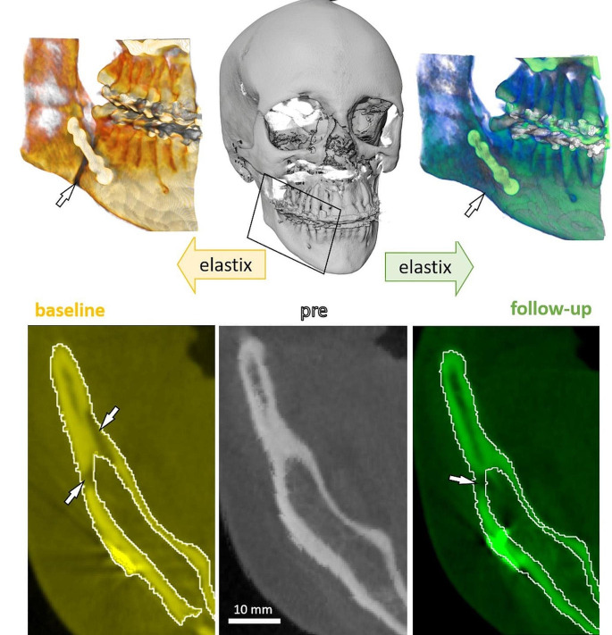 Figure 1
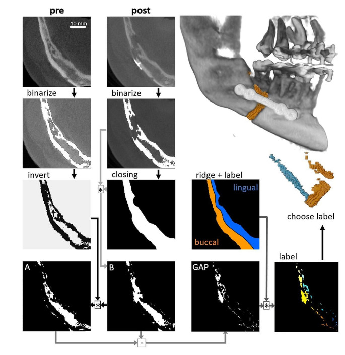 Figure 2
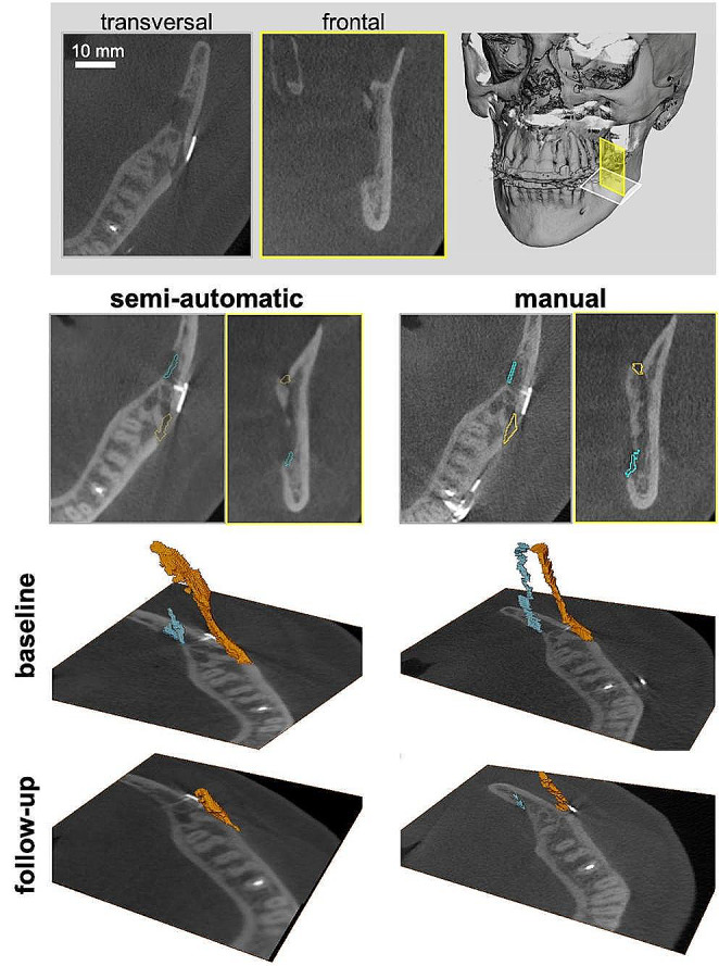 Figure 3
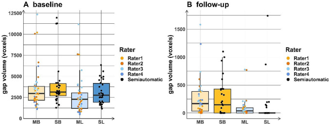 Figure 4
 Figure 5
Peer Reviews
No public reviews on file for this paper yet. If you reviewed it on a platform where reviews are public (OpenReview, ICLR, NeurIPS, ICML), you can paste yours below so the community can read it here.
Videos
No videos yet. Explain this paper in a talk, walkthrough, or lecture? Add one.
Taxonomy
TopicsDental Radiography and Imaging · Dental Implant Techniques and Outcomes · Orthodontics and Dentofacial Orthopedics