Cholangioscope-assisted endoscopic retrograde appendicitis therapy for the treatment of appendicitis complicated with fecal stones: a case report
Qi Zhao, Ruixia Wang, Maofeng Sun, Shulei Zhao

Abstract
Genes, proteins, chemicals, diseases, species, mutations and cell lines named across the full text — each resolved to its canonical identifier and authoritative record.
Click any figure to enlarge with its caption.
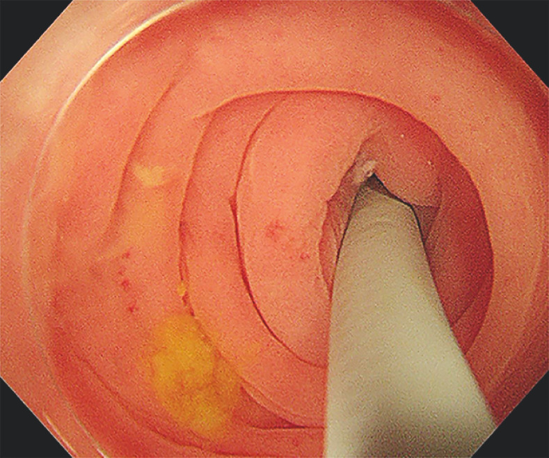 Fig. 1
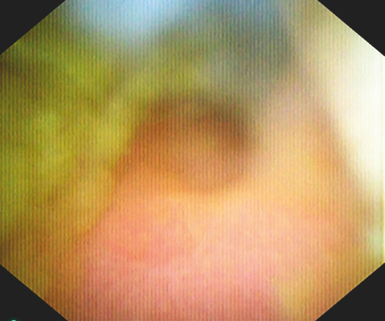 Fig. 2
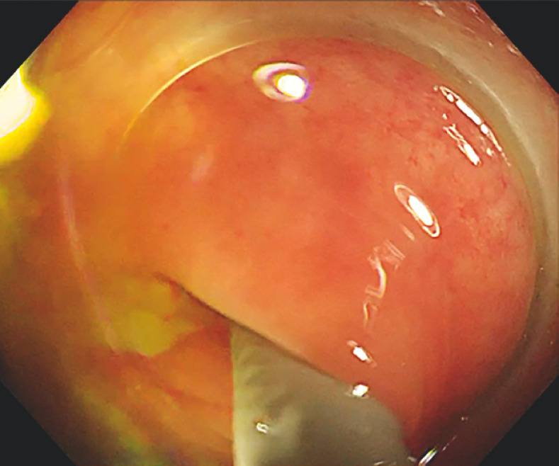 Fig. 3
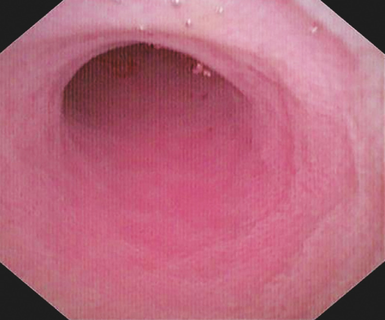 Fig. 4
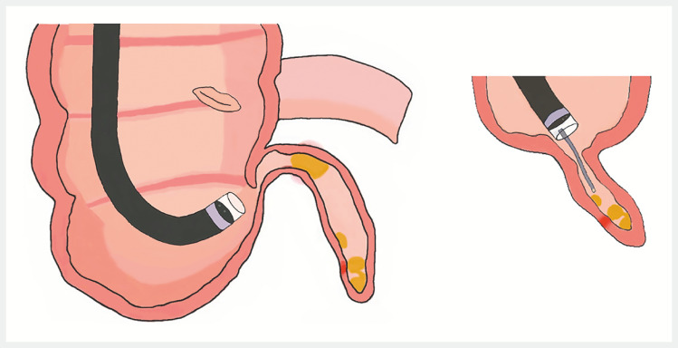 Fig. 5
Peer Reviews
No public reviews on file for this paper yet. If you reviewed it on a platform where reviews are public (OpenReview, ICLR, NeurIPS, ICML), you can paste yours below so the community can read it here.
Videos
No videos yet. Explain this paper in a talk, walkthrough, or lecture? Add one.
Taxonomy
TopicsAppendicitis Diagnosis and Management · Gallbladder and Bile Duct Disorders · Intestinal Malrotation and Obstruction Disorders
Acute appendicitis typically occurs when the opening of the appendix is blocked, most commonly by an impacted fecalith 1 . Surgical interventions for acute appendicitis often come with the drawbacks of increased postoperative complications and a high rate of removing the healthy appendix 2 . In 2012, a minimally invasive technique known as endoscopic retrograde appendicitis therapy (ERAT) was proposed. This method involves flushing out fecal stones and eliminating the blockage through the natural passage 3 . However, ERAT relies on X-ray and involves a complex procedure 4 . Drawing inspiration from the successful application of ultraslim endoscopes in treating complex bile duct stones, we incorporated a 9-Fr cholangioscope to assist with ERAT in managing appendicitis complicated with fecal stones. In this report, we present a case wherein this innovative approach was used.
A 15-year-old girl presented with recurrent right lower abdominal pain, leading to a diagnosis of acute appendicitis with fecal stones as revealed by ultrasound. Considering the patient's young age and her expressed wish to preserve her appendix, a decision was made to proceed with cholangioscope-assisted ERAT for treatment ( Video 1 ).
This video demonstrates the process of cholangioscope-assisted endoscopic retrograde appendicitis therapy for the treatment of appendicitis complicated with fecal stones.Video 1
A standard colonoscope with a 3.7-mm biopsy channel was used to reach the ileocecal region, where it was observed that the opening of the appendix appeared normal. Subsequently, a cholangioscope was inserted through the biopsy channel and successfully entered the appendix cavity ( Fig. 1 ). This allowed for the visualization of a significant amount of yellow fecal stones. With direct visual guidance from the cholangioscope, the fecal stones were effectively flushed out of the appendix cavity ( Fig. 2 ). After multiple flushing attempts ( Fig. 3 ), it was observed that the mucosa of the appendix cavity had become smooth and the lumen was no longer obstructed ( Fig. 4 ). The colonoscope and cholangioscope were then withdrawn. The pattern diagram of the treatment process for the appendicitis complicated with fecal stones in this case is shown below ( Fig. 5 ). Following the surgery, the patient experienced significant relief from the symptoms and signs. During a follow-up phone call 1 month later, the patient reported no abdominal pain.
Insertion of the cholangioscope into the opening of the appendix as observed during colonoscopy.
With direct visual guidance from the cholangioscope, the fecal stones were effectively flushed out of the appendix cavity.
After multiple flushing attempts, the fecal stones were flushed out of the appendix cavity under the colonoscope.
It was observed that the mucosa of the appendix cavity had become smooth and the lumen was no longer obstructed under the cholangioscope.
The pattern diagram of the treatment process for the appendicitis complicated with fecal stones in this case.
Compared to traditional ERAT, this operation does not rely on X-ray and provides a more comprehensive treatment for appendiceal fecal stones. The effectiveness and safety of ultraslim endoscope-assisted ERAT have been confirmed.
Endoscopy_UCTN_Code_TTT_1AQ_2AF
The reference list from the paper itself. Each links out to its DOI / PubMed record.
- 1Khan S Ali FS Ullah S Endoscopic retrograde appendicitis therapy: Is it really a need of the hour?Ann Surg 2023277 e 1e 435837901 10.1097/SLA.0000000000005576 · doi ↗ · pubmed ↗
- 2Yang B Kong L Ullah S Endoscopic retrograde appendicitis therapy versus laparoscopic appendectomy for uncomplicated acute appendicitis Endoscopy 20225474775410.1055/a-1737-638135021234 PMC 9329065 · doi ↗ · pubmed ↗
- 3Liu BR Song JT Han FY Endoscopic retrograde appendicitis therapy: a pilot minimally invasive technique (with videos)Gastrointest Endosc 20127686286622840292 10.1016/j.gie.2012.05.029 · doi ↗ · pubmed ↗
- 4Ullah S Ali FS Shi M Is it time for global adoption of endoscopic retrograde appendicitis therapy of acute appendicitis?Clin Res Hepatol Gastroenterol 20224610204910.1016/j.clinre.2022.10204936384200 · doi ↗ · pubmed ↗