Mammography of suspicious calcifications among ductal carcinoma in situ and benign breast disease
Wanrudee Lohitvisate, Chidsupang Kaeorat, Amolchaya Kwankua

TL;DR
The study compares suspicious calcifications in breast imaging to distinguish ductal carcinoma in situ from benign conditions, finding specific features that help early diagnosis.
Contribution
The study identifies linear morphology and segmental distribution as significant indicators of ductal carcinoma in situ (DCIS) in suspicious calcifications.
Findings
Linear morphology and segmental distribution of calcifications significantly correlate with DCIS.
Fine linear calcification increases the risk of DCIS with an odds ratio of 51.72.
Calcification descriptors are crucial for early diagnosis of DCIS versus benign breast disease.
Abstract
Most ductal carcinoma in situ (DCIS) lesions manifest early as calcifications, which could be benign or malignant. The classified group of suspicious calcifications among DCIS and benign breast disease is clinically important to early evaluate patient risk factors and plan treatment options. To compare imaging features of suspicious calcifications between DCIS and benign breast disease. A retrospective study of 101 suspicious calcifications was performed at Thammasat University Hospital from June 2011 to October 2020. The calcifications were surgically excised by mammography-guided wire localisation. The mammographic features of the suspicious calcifications were reviewed according to the fifth edition of the American College of Radiology Breast Imaging-Reporting and Data System lexicon. For comparing between two groups, the student t-test, Fisher’s exact test and Mann-Whitney U test…
Genes, proteins, chemicals, diseases, species, mutations and cell lines named across the full text — each resolved to its canonical identifier and authoritative record.
Click any figure to enlarge with its caption.
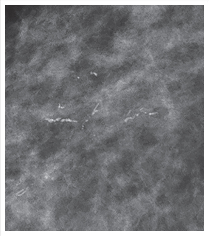 Figure 1
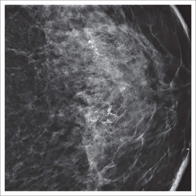 Figure 2
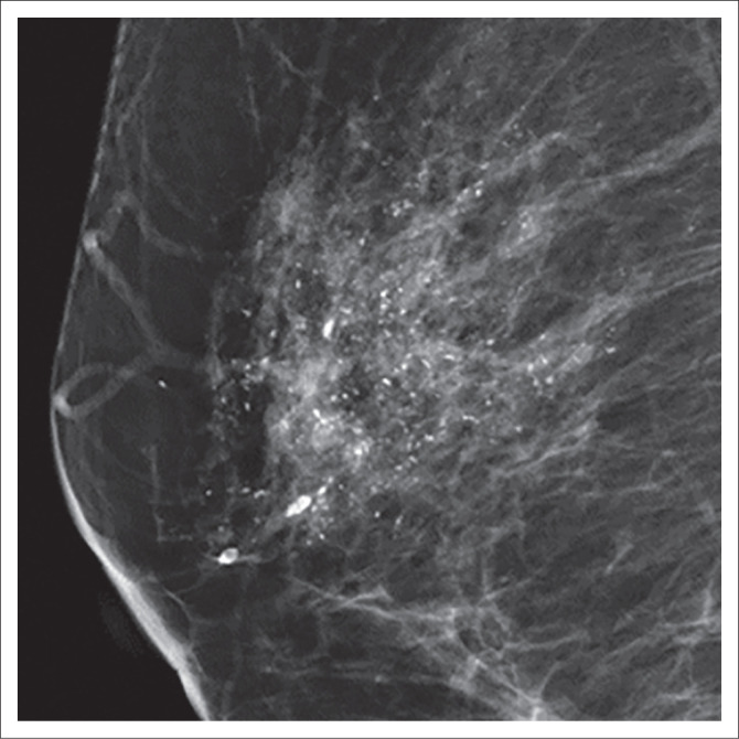 Figure 3
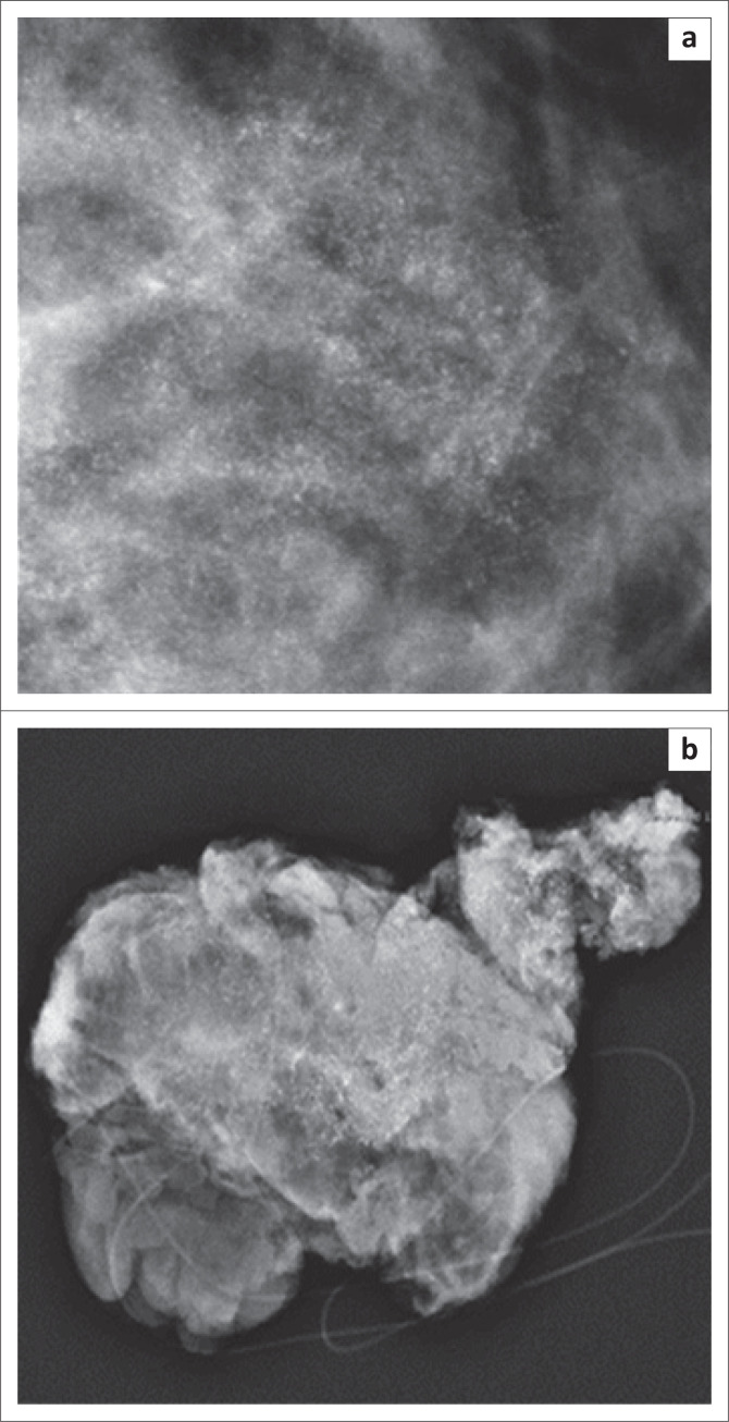 Figure 4
Peer Reviews
No public reviews on file for this paper yet. If you reviewed it on a platform where reviews are public (OpenReview, ICLR, NeurIPS, ICML), you can paste yours below so the community can read it here.
Videos
No videos yet. Explain this paper in a talk, walkthrough, or lecture? Add one.
Taxonomy
TopicsBreast Lesions and Carcinomas · Medical Imaging Techniques and Applications · Radiomics and Machine Learning in Medical Imaging