Correction: Dynein and Dynactin Leverage Their Bivalent Character to Form a High-Affinity Interaction
Amanda E. Siglin, Shangjin Sun, Jeffrey K. Moore, Sarah Tan, Martin Poenie, James D. Lear, Tatyana Polenova, John A. Cooper, John C. Williams

Abstract
Genes, proteins, chemicals, diseases, species, mutations and cell lines named across the full text — each resolved to its canonical identifier and authoritative record.
Click any figure to enlarge with its caption.
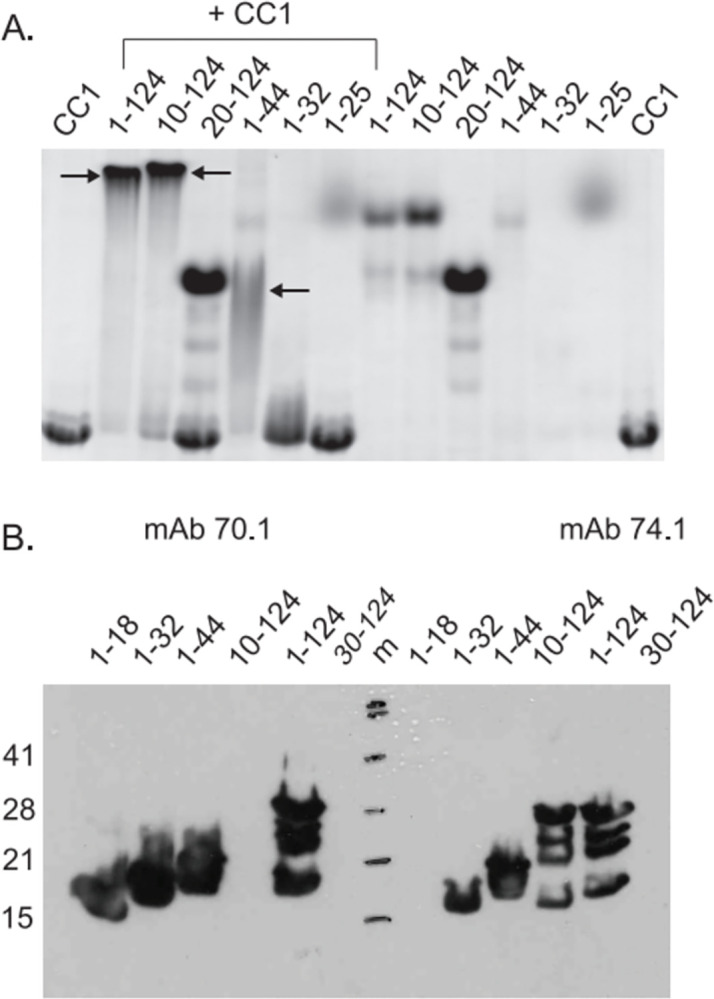 Figure 1
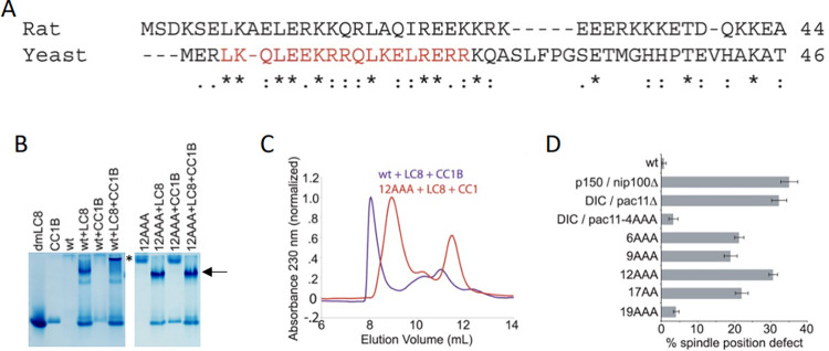 Figure 2
 Figure 3
Peer Reviews
No public reviews on file for this paper yet. If you reviewed it on a platform where reviews are public (OpenReview, ICLR, NeurIPS, ICML), you can paste yours below so the community can read it here.
Videos
No videos yet. Explain this paper in a talk, walkthrough, or lecture? Add one.
Taxonomy
TopicsMicrotubule and mitosis dynamics · Cellular transport and secretion · Photoreceptor and optogenetics research
This correction addresses errors in Figs 2, 3 and S3 of [1].
Specifically:
Regarding Fig 3D and S3B Fig in [1], the corresponding author stated that both figures were prepared from multiple gels as the full experiments did not fit on a single gel. These gels were then combined to produce Fig 3D and S3B Fig in [1].
Updated versions of Figs 2, 3, and S3 are provided with this Correction to address the above issues. The revised Fig 2B includes the correct original gel. Please note the updated Fig 2A and the corresponding labels have been inverted compared to the originally published Fig 2A in [1]. In the updated Fig 3 and S3 Fig, spaces have been added between lanes from distinct gels. Fig 3B and 3D and S3A and S3B Fig have been updated to match the figure legends for Fig 3 and S3 Fig.
The original uncropped images underlying the updated Figs 2, 3, and S3 are available in S1–8 Files provided with this notice.
The corresponding author stated that the individual-level quantitative data underlying this article [1] are no longer available.
The authors acknowledge, apologize, and are grateful for the opportunity to correct unintentional errors in the published article.
Supporting information
S3 FigAlanine scanning mutagenesis of Pac11 and statistical analysis of spindle position assays.(A) Native PAGE indicates that points mutants Pac11-L4A,K5A,Q6A, Pac11-Q6A,L7A,E8A, Pac11-E9A,K10A,R11A,Pac11-L17A,R18A, and Pac11-E19A,R20A,R21A abrogate Pac11-p150^Glued^ CC1B binding. In the presence of LC8, Pac11-p150^Glued^ CC1B binding is restored (indicated by an asterisk). Note only a slight change in migration is seen for the Pac11-p150^Glued^-LC8 complexes, however the CC1B band is absent or reduced indicating incorporation into the complex (arrow). Fig is composed of four separate native PAGE gels. (B) P-values were determined by t-test for mitotic spindle position assay. https://doi.org/10.1371/journal.pone.0059453.s003.(PDF)
S1 FileOriginal underlying image of labeled gel presented in Fig 2B.This file includes the original uncropped image used for Fig 2B on slide 1, and a repeat experiment, conducted at a later date, on slide 2.(PPTX)
S2 FileOriginal underlying images of labeled gels represented in Fig 3B and S3A Fig.This file includes the two original uncropped images used to compose the updated Fig 3B on slide 1, and the four original uncropped images used to compose the updated S3A Fig on slides 2 and 3.(PPTX)
S3 FileOriginal underlying image for gel presented in Fig 2B.(JPG)
S4 FileOriginal underlying image for gel presented in Fig 3B and S3A Fig. This file contains the original uncropped image for which lanes 3–6 correspond to lanes 7–10 of Fig 3B, and lanes 7–10 correspond to lanes 15–18 of S3A Fig.(PDF)
S5 FileOriginal underlying image for gel presented in Fig 3B and S3A Fig.This file contains the original uncropped image for which lanes 1–6 correspond to lanes 1–6 of Fig 3B and lanes 7–10 correspond to lanes 3–6 of S3A Fig.(PDF)
S6 FileOriginal underlying image for gel used in repeat experiment for data shown in Fig 3B and S3A Fig.This file contains the original uncropped image for a repeat experiment conducted at an earlier date. Lanes 1–6 are a repeat experiment of lanes 1–6 in Fig 3B and lanes 7–10 are a repeat experiment of lanes 3–6 in S3A Fig.(PDF)
S7 FileOriginal underlying image for gel presented in S3A Fig.This file contains the original uncropped image for which lanes 3–10 correspond to lanes 7–14 of S3A Fig.(PDF)
S8 FileOriginal underlying image for gel presented in S3A Fig.This file contains the original uncropped image for which lanes 1–10 correspond to lanes 1, 2 and 19–26 of S3A Fig.(PDF)
The reference list from the paper itself. Each links out to its DOI / PubMed record.