Quantitative assessment of left ventricular myocardial involvement in patients with connective tissue disease: a 3.0T contrast-enhanced cardiovascular magnetic resonance study
Jin Wang, Yue Gao, Zhi-Gang Yang, Ying-Kun Guo, Li Jiang, Rui Shi, Hua-Yan Xu, Shan Huang, Yuan Li

TL;DR
This study uses advanced heart imaging to show that patients with connective tissue diseases have reduced heart muscle function and blood flow, with more severe effects in non-IIM patients.
Contribution
The study introduces a quantitative CMR approach to assess myocardial involvement in CTD patients, revealing distinct differences between IIM and non-IIM subtypes.
Findings
CTD patients showed significantly reduced LV global myocardial deformation and microvascular perfusion compared to healthy controls.
Non-IIM patients exhibited lower LV global strain and longer time to maximum signal intensity compared to IIM patients.
Late gadolinium enhancement was independently associated with global radial and circumferential peak strain in CTD patients.
Abstract
The aim of this study was to evaluate left ventricular (LV) myocardial involvement in connective tissue disease (CTD) patients using multiparemetric imaging derived from cardiovascular magnetic resonance (CMR). CMR was performed on 146 CTD patients (comprising of 74 with idiopathic inflammatory myopathy (IIM) and 72 with non-IIM) and 72 healthy controls and included measures of LV global strains [including peak strain (PS), peak systolic (PSSR) and diastolic strain rate (PDSR)], myocardial perfusion [including upslope, max signal intensity (MaxSI), and time to maximum signal intensity (TTM)], and late gadolinium enhancement (LGE) parameters. Univariable and multivariable linear regression analyses were performed to determine the association between LV deformation and microvascular perfusion, as well as LGE. Our results indicated that CTD patients had decreased global longitudinal PS…
Genes, proteins, chemicals, diseases, species, mutations and cell lines named across the full text — each resolved to its canonical identifier and authoritative record.
Click any figure to enlarge with its caption.
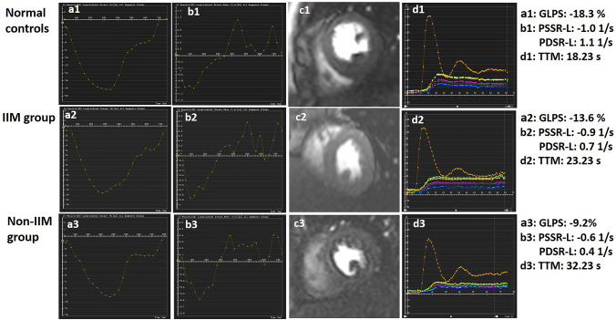 Figure 1
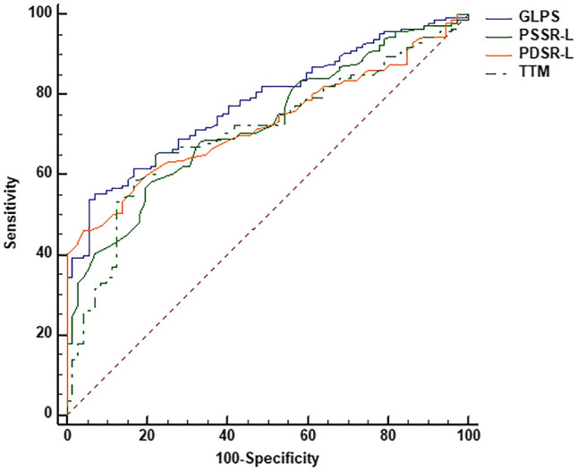 Figure 2
Peer Reviews
No public reviews on file for this paper yet. If you reviewed it on a platform where reviews are public (OpenReview, ICLR, NeurIPS, ICML), you can paste yours below so the community can read it here.
Videos
No videos yet. Explain this paper in a talk, walkthrough, or lecture? Add one.
Taxonomy
TopicsAdvanced MRI Techniques and Applications · Cardiac Imaging and Diagnostics · Cardiomyopathy and Myosin Studies