Oxidation–reduction imaging of myoglobin reveals two-phase oxidation in the reperfused myocardium
Sally Badawi, Clémence Leboullenger, Matthieu Chourrout, Yves Gouriou, Alexandre Paccalet, Bruno Pillot, Lionel Augeul, Radu Bolbos, Antonino Bongiovani, Nathan Mewton, Thomas Bochaton, Michel Ovize, Meryem Tardivel, Mazen Kurdi, Emmanuelle Canet-Soulas, Claire Crola Da Silva

TL;DR
This study uses myoglobin's oxidation states to track heart damage after blood flow is restored, offering a new imaging method for early myocardial infarction assessment.
Contribution
A novel imaging pipeline is developed to analyze myoglobin's redox states in the reperfused myocardium as a potential biomarker for myocardial infarction.
Findings
Myoglobin's fluorescence contributes to the myocardium's spectral signature under ischemia–reperfusion.
Oxidized myoglobin signal peaks 3 hours post-reperfusion and decreases with cardioprotection.
Early infarct size measured by oxidation–reduction imaging correlates with MRI results at 24 hours post-reperfusion.
Abstract
Myocardial infarction (MI) is a serious acute cardiovascular syndrome that causes myocardial injury due to blood flow obstruction to a specific myocardial area. Under ischemic–reperfusion settings, a burst of reactive oxygen species is generated, leading to redox imbalance that could be attributed to several molecules, including myoglobin. Myoglobin is dynamic and exhibits various oxidation–reduction states that have been an early subject of attention in the food industry, specifically for meat consumers. However, rarely if ever have the myoglobin optical properties been used to measure the severity of MI. In the current study, we develop a novel imaging pipeline that integrates tissue clearing, confocal and light sheet fluorescence microscopy, combined with imaging analysis, and processing tools to investigate and characterize the oxidation–reduction states of myoglobin in the ischemic…
Genes, proteins, chemicals, diseases, species, mutations and cell lines named across the full text — each resolved to its canonical identifier and authoritative record.
Click any figure to enlarge with its caption.
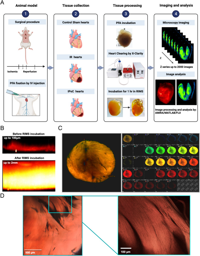 Figure 1
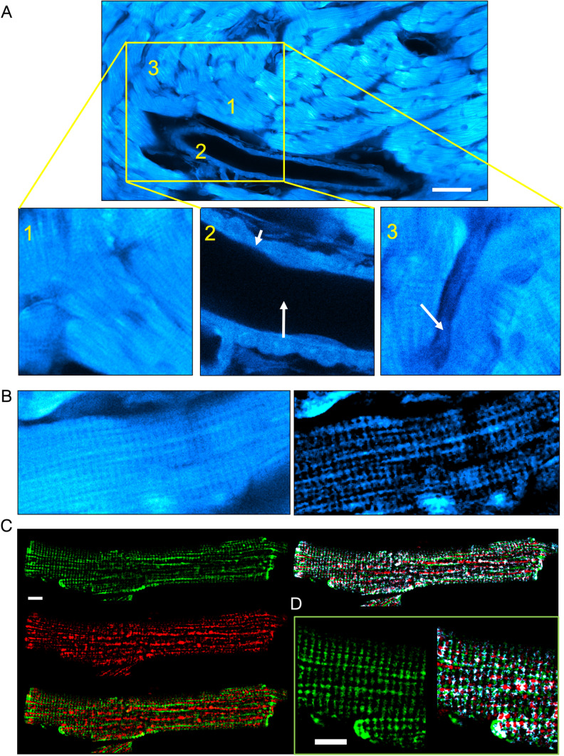 Figure 2
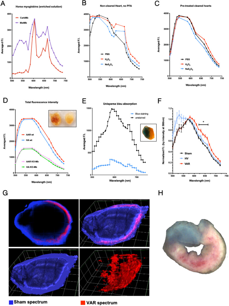 Figure 3
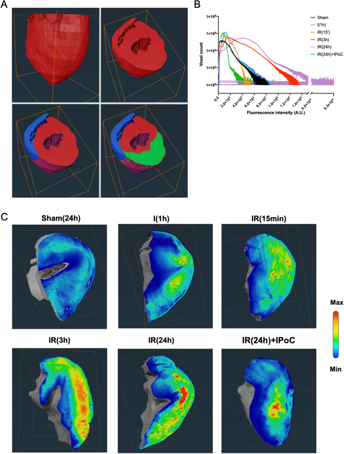 Figure 4
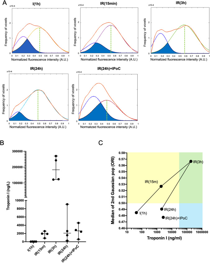 Figure 5
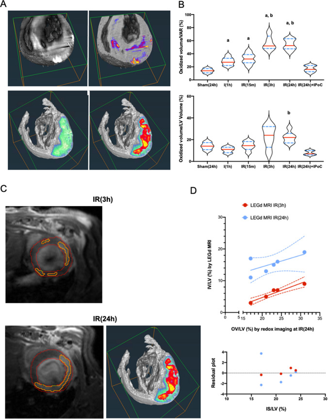 Figure 6
Peer Reviews
No public reviews on file for this paper yet. If you reviewed it on a platform where reviews are public (OpenReview, ICLR, NeurIPS, ICML), you can paste yours below so the community can read it here.
Videos
No videos yet. Explain this paper in a talk, walkthrough, or lecture? Add one.
Taxonomy
TopicsAdvanced MRI Techniques and Applications · Optical Imaging and Spectroscopy Techniques · Spectroscopy Techniques in Biomedical and Chemical Research