A 65‐year‐old woman with ALS and bilateral precentral motor band sign
Sadegh Ghaderi, Seyed Amir Hossein Batouli, Sanjay Kalra, Sana Mohammadi, Farzad Fatehi

TL;DR
This paper describes a case where advanced MRI techniques helped identify a specific brain sign in a woman with ALS, aiding early diagnosis.
Contribution
The paper highlights the use of QSM and other MRI techniques to detect the 'motor band sign' in ALS for the first time.
Findings
QSM MRI revealed a distinct 'motor band sign' in a patient with ALS.
The motor band sign indicates upper motor neuron involvement in ALS patients.
Abstract
Advanced MRI techniques, including SWI, MinIP, and QSM, are instrumental in detecting the “motor band sign” in ALS, aiding in the early diagnosis and assessment of upper motor neuron involvement, which is critical for therapeutic interventions. MRI with advanced techniques such as QSM reveals a distinct “motor band sign” in a patient with ALS, indicating upper motor neuron involvement.
Genes, proteins, chemicals, diseases, species, mutations and cell lines named across the full text — each resolved to its canonical identifier and authoritative record.
Click any figure to enlarge with its caption.
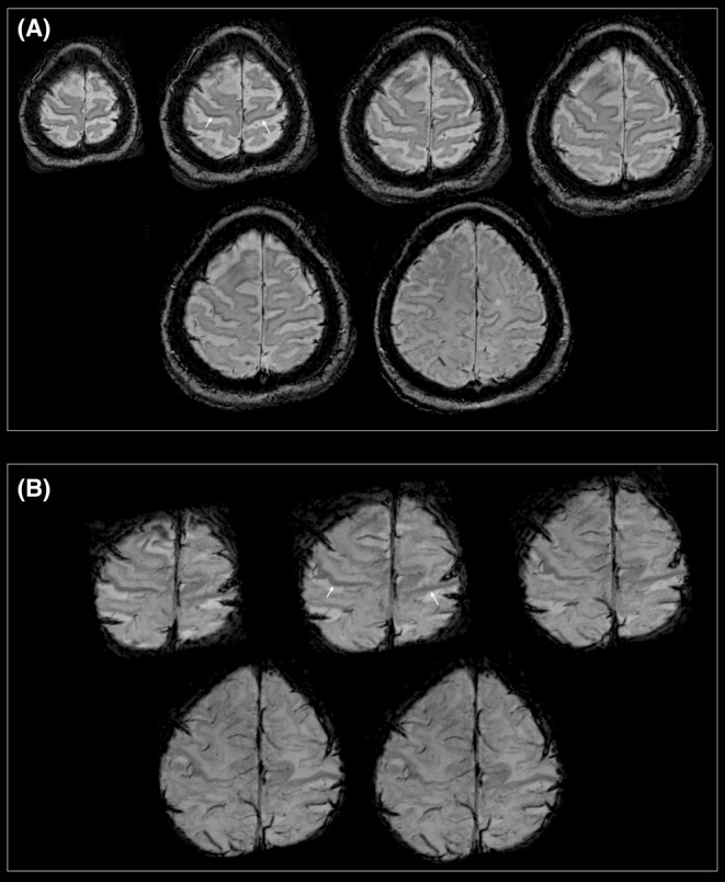 FIGURE 1
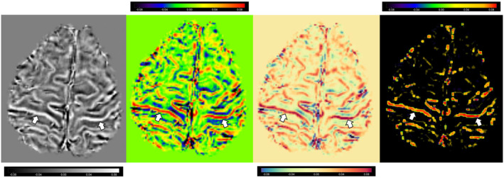 FIGURE 2
Peer Reviews
No public reviews on file for this paper yet. If you reviewed it on a platform where reviews are public (OpenReview, ICLR, NeurIPS, ICML), you can paste yours below so the community can read it here.
Videos
No videos yet. Explain this paper in a talk, walkthrough, or lecture? Add one.
Taxonomy
TopicsAmyotrophic Lateral Sclerosis Research · Neurological disorders and treatments · Genetic Neurodegenerative Diseases
CLINICAL STUDY
1
Case presentation and imaging
1.1
A 65‐year‐old woman who was illiterate and worked as a housewife and gardener was referred to the amyotrophic lateral sclerosis (ALS) clinic. She was diagnosed with clinically definite limb‐onset ALS using the Awaji criteria. The initial symptoms appeared 31 months prior to imaging. At the time of imaging, her ALSFRS‐R and BMI scores were 43 and 42.44 kg/m^2^, respectively. Imaging, including susceptibility‐weighted imaging (SWI) and minimum intensity projection (MinIP) protocols and acquisitions, as well as post‐processing for quantitative susceptibility mapping (QSM) analyses, was conducted based on our previous study.1 SWI visually displays tissue magnetic field variations using magnitude and phase information, whereas QSM quantifies the underlying magnetic susceptibilities that cause these variations.1 However, in this study, we used a visual map of the susceptibility to precentral MBS.
RESULTS AND DISCUSSION
2
MRI revealed a bilateral decreased signal (hypointensity) on SWI, MinIP, and QSM images, consistent along the precentral gyri, which is referred to as the “motor band sign” or “black ribbon sign”1, 2 (Figures 1 and 2). The precentral motor band sign is a suggestive and sensitive biomarker for motor neuron diseases (MNDs) such as ALS1 and other neurological disorders such as Huntington's disease.3 This sign is believed to result from the accumulation of iron in the motor cortex owing to oxidative stress and microglia‐driven neuroinflammation.1 MRI with MinIP and/or SWI and post‐processing techniques such as QSM may play a vital role in the diagnosis of rare cases of ALS and in assessing UMN involvement in therapeutic trials.1, 2 Thus, advanced MRIs, such as T2*‐weighted, transverse relaxation rates, SWI, and QSM, have been proposed to detect radiological markers of upper motor neuron degeneration (UMN) in ALS.1, 2
Motor band signs (white arrows) showing bilateral hypointense signals in the precentral region on (A) susceptibility‐weighted imaging (SWI) and (B) minimum intensity projection (MinIP) images in a patient diagnosed with ALS.
Quantitative susceptibility mapping images (using four different filters with MRIcroGL) of the bilateral precentral gyrus showing motor‐band signs (white arrows).
Close monitoring for emerging MND should be considered when MBS is present on the SWI/QSM. In research and clinical settings, SWI and QSM are useful for detecting cortical neuroimaging biomarkers of UMN degeneration in MNDs.1, 2 By combining structural, functional, metabolic, and quantitative techniques, multimodal MRI can provide the best imaging biomarkers. However, feasible protocols for clinical use must be optimized.
AUTHOR CONTRIBUTIONS
Sadegh Ghaderi: Conceptualization; data curation; formal analysis; investigation; methodology; project administration; resources; software; validation; visualization; writing – original draft; writing – review and editing. Seyed Amir Hossein Batouli: Conceptualization; investigation; project administration; validation; visualization; writing – review and editing. Sanjay Kalra: Conceptualization; investigation; project administration; validation; visualization; writing – review and editing. Sana Mohammadi: Data curation; formal analysis; investigation; methodology; resources; software; validation; writing – original draft; writing – review and editing. Farzad Fatehi: Conceptualization; investigation; methodology; project administration; supervision; validation; visualization; writing – original draft; writing – review and editing.
FUNDING INFORMATION
3
This research received no specific grant from any funding agency in the public, commercial, or not‐for‐profit sectors.
CONFLICT OF INTEREST STATEMENT
The authors declare that they have no conflict of interest.
ETHICAL STATEMENT
The study was approved by the Ethics Committee of the Tehran University of Medical Sciences (Ethical Code: IR.TUMS.MEDICINE.REC.1400.1173). We obtained written consent from our patient, who may have an identifiable image or data.
CONSENT
Written informed consent was obtained from the patient to publish this report in accordance with the journal's patient consent policy.
The reference list from the paper itself. Each links out to its DOI / PubMed record.
- 1Mohammadi S , Ghaderi S . Motor band sign in motor neuron diseases using magnetic resonance imaging: a systematic review. Acta Neurol Scand. 2023;2023:e 6677967. doi:10.1155/2023/6677967 · doi ↗
- 2Krieger C , Kalra S . Imaging the amyotrophic lateral sclerosis brain: the motor band sign. Can J Neurol Sci. 2023;50(3):327‐328. doi:10.1017/cjn.2022.67 35634738 · doi ↗ · pubmed ↗
- 3Dash D , Mestre TA . Motor band sign in a Huntington disease phenocopy. Parkinsonism Relat Disord. 2023;109:105333. doi:10.1016/j.parkreldis.2023.105333 36854213 · doi ↗ · pubmed ↗