Improving explanation of motor disability with diffusion-based graph metrics at onset of the first demyelinating event
Michael A Foster, Ferran Prados, Sara Collorone, Baris Kanber, Niamh Cawley, Indran Davagnanam, Marios C Yiannakas, Lola Ogunbowale, Ailbhe Burke, Frederik Barkhof, Claudia AM Gandini Wheeler-Kingshott, Olga Ciccarelli, Wallace Brownlee, Ahmed T Toosy

TL;DR
This study shows that graph metrics from brain scans can better explain motor disability in early multiple sclerosis than traditional MRI methods.
Contribution
The study introduces diffusion-based graph metrics as a novel way to assess motor disability in early multiple sclerosis.
Findings
Graph metrics like local efficiency, clustering, and transitivity were reduced in patients compared to controls.
Higher assortativity in brain networks was linked to higher disability scores and faster walking times.
Adding graph metrics to conventional MRI improved prediction of disability outcomes.
Abstract
Conventional magnetic resonance imaging (MRI) does not account for all disability in multiple sclerosis. The objective was to assess the ability of graph metrics from diffusion-based structural connectomes to explain motor function beyond conventional MRI in early demyelinating clinically isolated syndrome (CIS). A total of 73 people with CIS underwent conventional MRI, diffusion-weighted imaging and clinical assessment within 3 months from onset. A total of 28 healthy controls underwent MRI. Structural connectomes were produced. Differences between patients and controls were explored; clinical associations were assessed in patients. Linear regression models were compared to establish relevance of graph metrics over conventional MRI. Local efficiency (p = 0.045), clustering (p = 0.034) and transitivity (p = 0.036) were reduced in patients. Higher assortativity was associated with…
Genes, proteins, chemicals, diseases, species, mutations and cell lines named across the full text — each resolved to its canonical identifier and authoritative record.
Click any figure to enlarge with its caption.
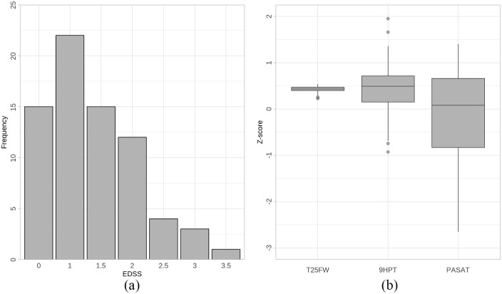 Figure 1
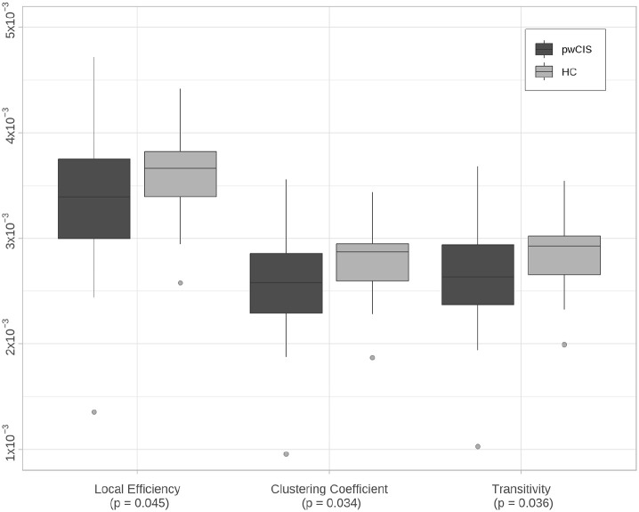 Figure 2
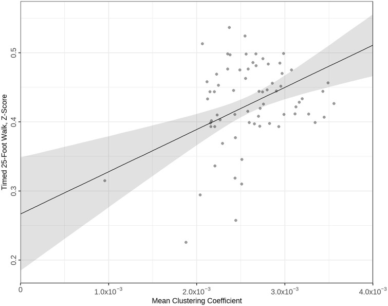 Figure 3
 Figure 4
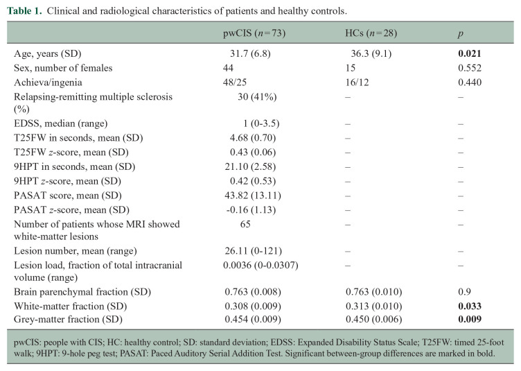 Figure 5
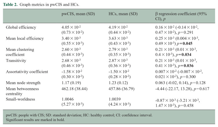 Figure 6
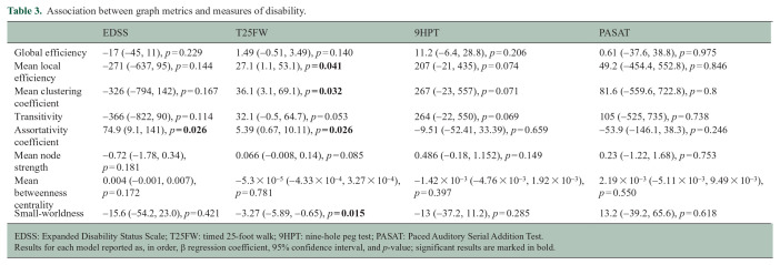 Figure 7
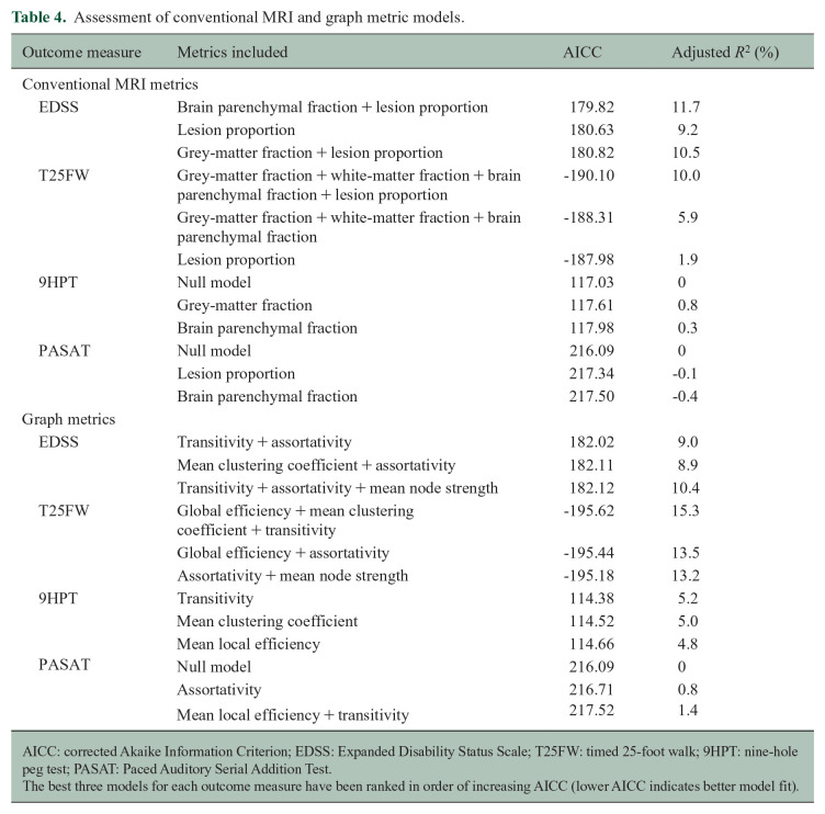 Figure 8
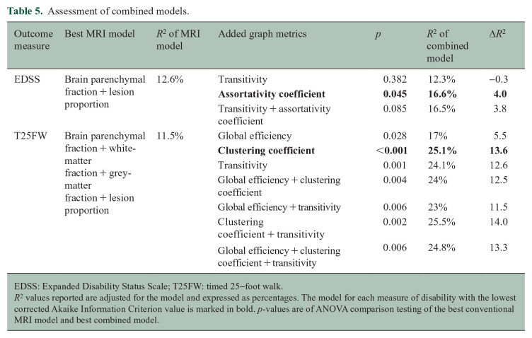 Figure 9
Peer Reviews
No public reviews on file for this paper yet. If you reviewed it on a platform where reviews are public (OpenReview, ICLR, NeurIPS, ICML), you can paste yours below so the community can read it here.
Videos
No videos yet. Explain this paper in a talk, walkthrough, or lecture? Add one.
Taxonomy
TopicsAdvanced Neuroimaging Techniques and Applications · Functional Brain Connectivity Studies · Neonatal and fetal brain pathology