Unmasking the Mimic: Radionecrotic Lesion Masquerading as Brain Neoplasia on Magnetic Resonance Imaging
Diego A. Barrios-González, Jimena Gonzalez-Salido, Jimena Colado-Martínez, Santiago Philibert-Rosas, Fernando Sotelo-Díaz, Mario A. Sebastián-Díaz, L. Jimena Gómez-Rodríguez, Nora E. Kerik-Rotenberg, Guillermo A. Gutiérrez-Aceves, Iris E. Martínez-Juárez.

TL;DR
A patient who underwent brain radiosurgery developed a lesion that looked like a tumor on MRI, but was actually radionecrosis confirmed using advanced imaging techniques.
Contribution
This paper presents a unique case of radionecrosis mimicking a brain tumor after radiosurgical callosotomy, emphasizing the need for accurate differential diagnosis.
Findings
Radionecrosis can mimic CNS tumors on MRI, requiring advanced imaging for differentiation.
18-FDG PET/CT and 11C-acetate PET/CT scans helped distinguish radionecrosis from neoplasia.
The case highlights the importance of considering radionecrosis in post-radiosurgery patients with suspicious lesions.
Abstract
Corpus callosotomy is a therapeutic approach for drug-resistant epilepsy, with positive outcomes observed in managing atonic seizures. Despite a decline in its usage, radiosurgical callosotomy remains a viable option for drug-resistant epilepsy due to its low risks of post-radiation neoplasia, albeit not with exceptions. Brain radionecrosis is characterized by tissue death and vascular endothelial damage following the procedure. Despite the low risk of intracranial secondary malignancy associated with radiation in some cases, post-radiation lesions might present with distinct characteristics needing a thorough diagnostic approach. Herein, we present a unique case of a patient with focal epilepsy who developed a radionecrotic lesion following radiosurgical callosotomy, affecting the anterior cingulate cortex, and mimicking a central nervous system (CNS) tumor. Molecular imaging…
Genes, proteins, chemicals, diseases, species, mutations and cell lines named across the full text — each resolved to its canonical identifier and authoritative record.
Click any figure to enlarge with its caption.
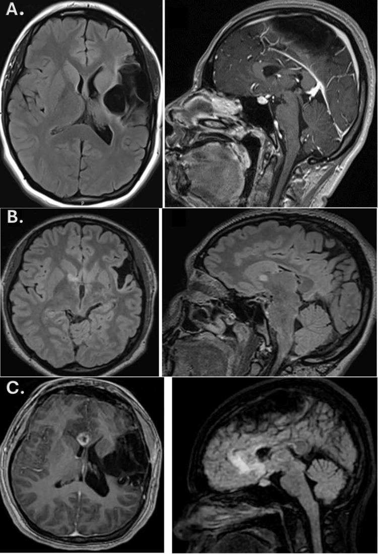 Figure 1
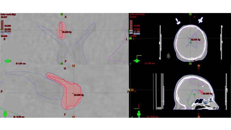 Figure 2
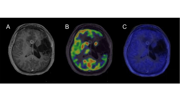 Figure 3
Peer Reviews
No public reviews on file for this paper yet. If you reviewed it on a platform where reviews are public (OpenReview, ICLR, NeurIPS, ICML), you can paste yours below so the community can read it here.
Videos
No videos yet. Explain this paper in a talk, walkthrough, or lecture? Add one.
Taxonomy
TopicsGlioma Diagnosis and Treatment · Brain Metastases and Treatment · Cerebrospinal fluid and hydrocephalus