Ultrasound image-based nomogram combining clinical, radiomics, and deep transfer learning features for automatic classification of ovarian masses according to O-RADS
Lu Liu, Wenjun Cai, Hongyan Tian, Beibei Wu, Jing Zhang, Ting Wang, Yi Hao, Guanghui Yue

TL;DR
This study creates a tool that combines ultrasound images and clinical data to help classify ovarian masses as benign or malignant, improving accuracy and supporting junior radiologists.
Contribution
A novel nomogram integrating clinical, radiomics, and deep learning features for automatic ovarian mass classification according to O-RADS.
Findings
The nomogram achieved an AUC of 0.930 and outperformed junior radiologists in diagnostic accuracy.
Junior radiologists' performance significantly improved with the model's assistance.
The model showed good calibration and clinical utility in decision curve analysis.
Abstract
Accurate and rapid discrimination between benign and malignant ovarian masses is crucial for optimal patient management. This study aimed to establish an ultrasound image-based nomogram combining clinical, radiomics, and deep transfer learning features to automatically classify the ovarian masses into low risk and intermediate-high risk of malignancy lesions according to the Ovarian- Adnexal Reporting and Data System (O-RADS). The ultrasound images of 1,080 patients with 1,080 ovarian masses were included. The training cohort consisting of 683 patients was collected at the South China Hospital of Shenzhen University, and the test cohort consisting of 397 patients was collected at the Shenzhen University General Hospital. The workflow included image segmentation, feature extraction, feature selection, and model construction. The pre-trained Resnet-101 model achieved the best…
Genes, proteins, chemicals, diseases, species, mutations and cell lines named across the full text — each resolved to its canonical identifier and authoritative record.
Click any figure to enlarge with its caption.
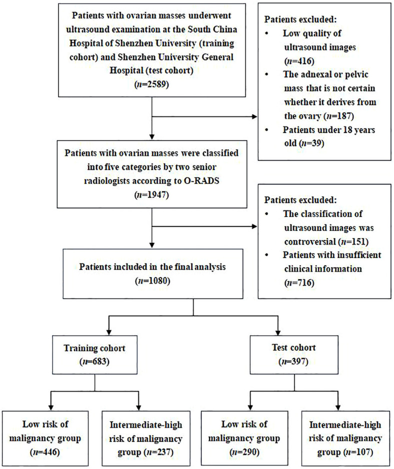 Figure 1
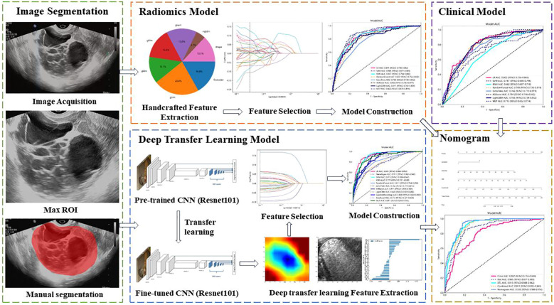 Figure 2
 Figure 3
 Figure 4
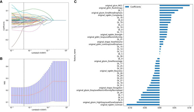 Figure 5
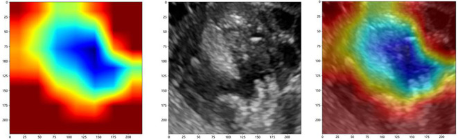 Figure 6
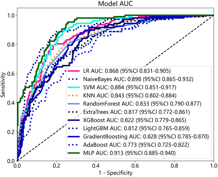 Figure 7
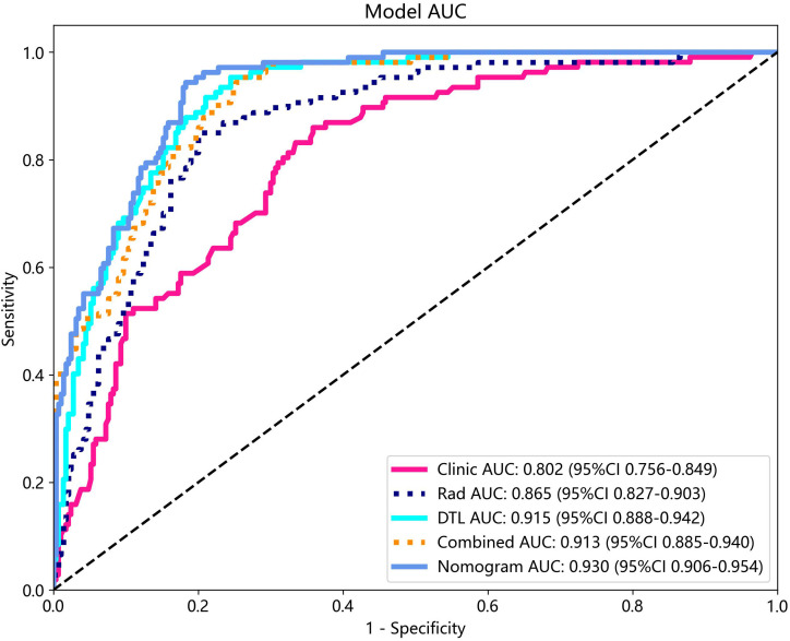 Figure 8
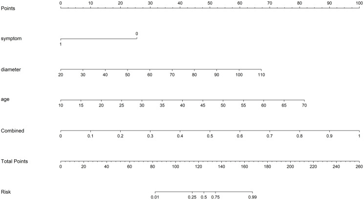 Figure 9
 Figure 10
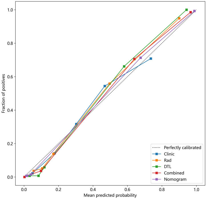 Figure 11
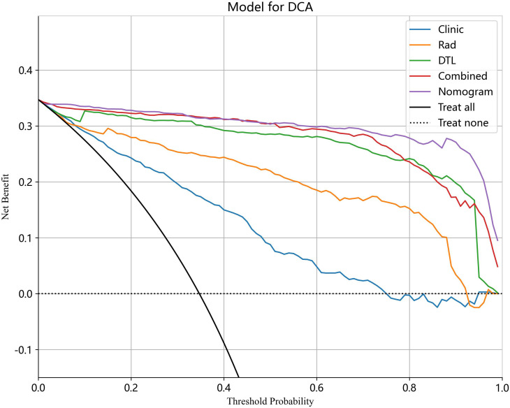 Figure 12
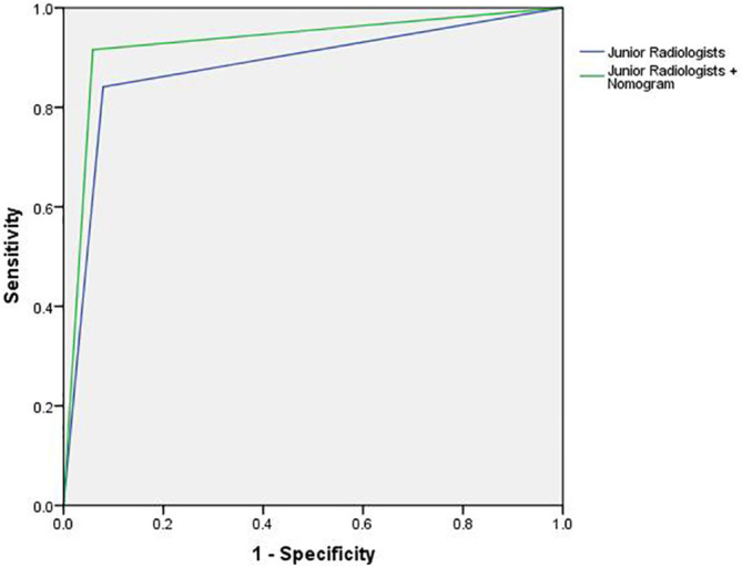 Figure 13
Peer Reviews
No public reviews on file for this paper yet. If you reviewed it on a platform where reviews are public (OpenReview, ICLR, NeurIPS, ICML), you can paste yours below so the community can read it here.
Videos
No videos yet. Explain this paper in a talk, walkthrough, or lecture? Add one.
Taxonomy
TopicsRadiology practices and education · Ovarian cancer diagnosis and treatment · Ectopic Pregnancy Diagnosis and Management