Clinical Case of Mild Tatton–Brown–Rahman Syndrome Caused by a Nonsense Variant in DNMT3A Gene
Fatima Bostanova, Olga Levchenko, Margarita Sharova, Natalia Semenova

TL;DR
A 10-year-old boy with a rare genetic disorder caused by a new mutation in the DNMT3A gene shows mild symptoms, expanding the known range of this condition.
Contribution
This is the first report of a recurrent nonsense variant in DNMT3A, suggesting a mutational hot-spot and highlighting a milder clinical presentation.
Findings
A de novo nonsense variant in DNMT3A (c.1443C>A, p.Tyr481Ter) was identified in a patient with mild Tatton–Brown–Rahman syndrome.
The case expands the phenotypic spectrum of the syndrome, showing milder symptoms than typically reported.
The variant is a potential mutational hot-spot, as it is recurrent and not found in the patient's parents.
Abstract
Tatton–Brown–Rahman syndrome is a rare autosomal dominant hereditary disease caused by pathogenic variants in the DNMT3A gene, which is an important participant in epigenetic regulation, especially during embryonic development, and is highly expressed in all tissues. The main features of the syndrome are high growth, macrocephaly, intellectual disability, and facial dysmorphic features. We present a clinical case of Tatton–Brown–Rahman syndrome in a ten-year-old boy with macrocephaly with learning difficulties, progressive eye impairment, and fatigue suspected by a deep learning-based diagnosis assistance system, Face2Gene. The proband underwent whole-exome sequencing, which revealed a recurrent nonsense variant in the 12th exon of the DNMT3A, leading to the formation of a premature stop codon—NM_022552.5:c.1443C>A (p.Tyr481Ter), in a heterozygous state. This variant was not found in…
Genes, proteins, chemicals, diseases, species, mutations and cell lines named across the full text — each resolved to its canonical identifier and authoritative record.
Click any figure to enlarge with its caption.
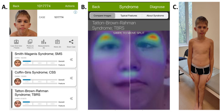 Figure 1
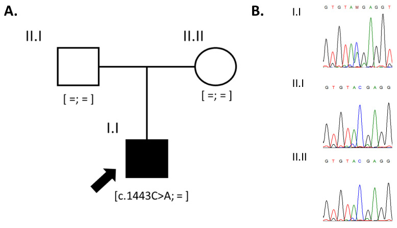 Figure 2
- —Ministry of Science and Higher Education of the Russian Federation
Peer Reviews
No public reviews on file for this paper yet. If you reviewed it on a platform where reviews are public (OpenReview, ICLR, NeurIPS, ICML), you can paste yours below so the community can read it here.
Videos
No videos yet. Explain this paper in a talk, walkthrough, or lecture? Add one.
Taxonomy
TopicsRNA modifications and cancer · Cancer-related molecular mechanisms research · Cancer-related gene regulation
1. Introduction
Tatton–Brown–Rahman syndrome (TBRS) is caused by a heterozygous causative variant in the DNMT3A gene [1]. Its hallmark clinical features include postnatal macrosomia, intellectual disability, and distinct facial dysmorphisms. Its expected prevalence is <1/1,000,000. The gene’s protein product, DNA methyltransferase 3A, operates in DNA modification via methylation [2].
Our understanding of the pathogenesis of this syndrome remains incomplete. In 2018, Tatton–Brown et al. published an article presenting a comprehensive characterization of 55 TBRS patients, revealing consistent phenotypic traits, including the following: tall stature, macrocephaly, dense horizontal eyebrows, narrow eye slits, an elongated oval face, a high-arched palate, and varying degrees of intellectual disability. Some patients also displayed additional features with variable frequency, such as mitral and tricuspid valve defects, atrial septal defects, pronounced joint hypermobility, and umbilical hernia [3,4].
In 2017, the first documented case of TBRS associated with the p.Arg882Cys missense substitution was reported. The patient developed acute myeloid leukemia (AML) at age 15 [5]. Notably, this single-nucleotide variant (SNV) also stands as the most common somatic mutation in AML [6,7,8,9]. Concurrently, Shen et al. established the association of eight additional SNVs found in TBRS with AML [10].
Notably, beyond its association with TBRS and AML, germline mutations within the DNMT3A gene could lead to Gaine–Spratou–Jackson syndrome (GSJS), characterized by a clinical phenotype. In stark contrast to TBRS, GSJS manifests with features such as dwarfism, low weight, microcephaly, and intellectual disability. Gaine et al., in their study, not only provided clinical descriptions of patients but also conducted a functional analysis of identified missense variants (p.Trp330Arg and Asp333Asn). Their investigation revealed an association between these variants and hypermethylation across diverse genomic regions, classifying them as gain-of-function (GoF) variants [11]. Considering the presence of both loss-of-function variants and GoF in the TBRS gene, it is reasonable to hypothesize that the variant’s impact on protein function contributes to the divergent phenotype. However, comprehensive investigations are essential to solidify and establish robust genotype–phenotype correlations.
2. Materials and Methods
Subjects: The clinical examination and genetic analysis of the proband was performed in the Research Centre for Medical Genetics, Russia. All research participants gave their informed consent to the clinical examination and publication of their anonymized data (for infant probands, the adult responsible for them signed a consent form). The study was performed in accordance with the Declaration of Helsinki and approved by the Institutional Review Board of the Research Centre for Medical Genetics, Russia. Written informed consent was obtained from the family.
Genetic analysis: The manuscript RefSeq accession numbers NG_029465.2, NM_022552.5, and NP_072046.2 were used for the DNMT3A gene.
Whole-exome sequencing (WES) of genomic DNA was performed on an Illumina NextSeq 500 instrument (Illumina, San Diego, USA) in 2 × 151 bp paired-end mode to an average depth of minimum 70×. The libraries were prepared and enriched using Illumina Nextera Rapid Capture Exome Kit v1.2.
Sanger sequencing was performed using the ABI PRISM Big Dye Terminator (v 3.1) Cycle Sequencing Kit (Applied Biosystems, Foster City, CA, USA) on the ABI3130xl Genetic Analyzer (Applied Biosystems, Foster City, CA, USA) according to the manufacturer’s recommendations.
3. Results
3.1. Case Description
The proband, a 10-year-old boy from a non-consanguineous family, received medical consultation in our center. The family pedigree was unremarkable, with two healthy elder sons. The proband was born from the mother’s third pregnancy with a birth weight of 4950 g (+2.83 SD), length of 60 cm (+3.75 SD), and an Apgar score of 8/9. Motor development occurred with minor temporal delays: he started to hold his head at 4 months, sat independently at 7 months, and walked independently at the age of 1 year and 3 months. Speech development initially matched age expectations, with a vocabulary of approximately ten words by the age of one. However, phrase speech emerged only at 3.5 years. The proband has been under the care of a neuropsychologist and a neurologist since the age of 3 years. Currently, he attends a specialized school with average academic performance.
Magnetic resonance imaging (MRI) of the brain conducted at the age of 4 revealed no signs of structural or focal pathology. Electroencephalography (EEG) showed no epileptiform activity. Echocardiography identified mitral and tricuspid valve prolapses. Progressive visual impairment was noted by the age of 8.5. Ophthalmological evaluation revealed partial atrophy of the optic nerves, secondary convergent strabismus, and simple myopic astigmatism in both eyes.
According to a medical psychologist consultation, the child exhibited a diminished intellectual capacity with immaturity and specific characteristics in the formation of the emotional–volitional sphere (according to the Wechsler scale: Verbal Intellectual Quotient (VIQ)—80, Non-Verbal Intellectual Quotient (NIQ)—85, Overall Intellectual Quotient (OI)—80). Speech–language pathology of a mild degree, dysgraphia, and elements of dyslexia were observed, leading to ongoing observation by a speech therapist due to systemic speech underdevelopment. There were no autism spectrum disorder features. Endocrine etiology for the child’s excessive growth was excluded through comprehensive examination (growth hormone: 6.4 ng/mL, IGF-1: 201 ng/mL, Prolactin: 259 mME/m). Also, he had no history of any oncological conditions.
Upon objective examination at the age of 10, the proband’s height was 146 cm (+1.10 SD), with a weight of 44 kg (+1.32 SD) and head circumference of 56 cm (+2.23 SD). Some dysmorphic features were noted, including downslanted palpebral fissures; epicanthic fold; convergent strabismus; large, dysplastic, posteriorly rotated ears; a thin upper lip; prominent incisors; and a crowded dental arch (Figure 1A,C). Generalized joint hypermobility was observed. Neurological examination revealed no focal neurological symptoms. No seizures were reported, and the child’s behavior was calm, showing an ability to comprehend directed speech and follow simple instructions. An analysis of the proband’s facial phenotype using a deep learning-based diagnosis assistance system, Face2Gene, did not reveal any high-gestalt match (Figure 1A,B), but TBRS was listed third according to the analysis.
3.2. Genetic Analysis
Cytogenetic analysis was conducted on the child with developmental delay, revealing a normal male karyotype of 46, XY. Targeted genetic testing for Martin–Bell syndrome and Beckwith–Wiedemann syndrome was performed using molecular genetic methods, revealing no pathological findings.
Whole-exome sequencing revealed a previously reported [12] variant in exon 12 in the DNMT3A gene (hg19—chr2:25468920G>T) in a heterozygous state. This variant is absent in the gnomAD database and leads to the formation of a premature stop codon—c.1443C>A (p.Tyr481Ter). Segregation analysis of the identified variant (p.Tyr481Ter, NM_175629) in the DNMT3A gene confirmed its de novo status (Figure 2B), and based on the ACMG guidelines [13], it was classified as pathogenic (PM2, PVS1, PS2). Heterozygous pathogenic variants in the DNMT3A gene are associated with Tatton–Brown–Rahman syndrome, which was confirmed based on the results of the genetic test of the proband.
4. Discussion
Our proband presented a mild manifestation of TBRS, prompting a reasonable comparison of their clinical profile with other macrosomic syndromes. Distinguishing these syndromes clinically is feasible and highly important for ensuring the right diagnostic journey. We want to emphasize the importance of using Face2Gene even in patients with mild phenotypes to optimize patients’ diagnostic journey.
In our proband, various traits reminiscent of Sotos syndrome were observed, including accelerated growth alongside normal endocrine function, developmental delays in psychological and motor skills, and behavioral abnormalities featuring elements of aggression and learning difficulties. Notably absent were distinct neurological markers typically associated with Sotos syndrome, such as ataxia, tremor, coordination disturbances, and epileptic activity. It is essential to highlight the divergent growth patterns—Sotos syndrome showcases accelerated growth concurrent with a deficit in body mass, while TBRS patients commonly exhibit obesity. Our patient displayed a trend of rapid weight gain, leading to adherence to a controlled diet over the past 5 months, resulting in a weight reduction of 20 kg. Specific dysmorphic features characteristic of Sotos syndrome, such as dolichocephaly, prominent frontal bosses, macroglossia, and a triangular face, were not observed in our proband [14].
Weaver syndrome is another syndrome that enables one to make a diagnosis different from TBRS. Like TBRS and Sotos syndrome, Weaver syndrome is characterized by the presence of pre- and postnatal overgrowth and variable intellectual disability. However, specific facial features, such as a broad forehead, widely spaced eyes, and almond-shaped palpebral fissures, in combination with a hoarse, low-pitched cry in infants; camptodactyly; and doughy skin, can help to distinguish it from previously described syndromes [15].
Also, both pre- and postnatal macrosomia were noted in our patient, as observed in Beckwith–Wiedemann syndrome (BWS). However, distinctive clinical features, such as visceromegaly, macroglossia, omphalocele, and neonatal hypoglycemia, characteristic of BWS were absent in our proband. It is worth noting that developmental delay is not a typical feature of Beckwith–Wiedemann syndrome [16]. We want to emphasize the specific molecular mechanisms of Beckwith–Wiedemann syndrome, like the abnormal methylation pattern at 11p15.5 or copy number variant of chromosome 11p15.5, which could not be detected by single-gene or multigene NGS panels testing, unlike with TBRS, Sotos syndrome, and Weaver syndrome.
Distinguishing TBRS from Cowden syndrome is facilitated by the absence of multiple hamartomas. However, discerning between the allelic variant of Cowden syndrome—macrocephaly and autism syndrome—and TBRS is more complex. Our patient exhibits partial optic nerve atrophy, a feature not typical of other syndromes involving macrocephaly or excessive growth [17].
Considering the patient’s medical history, including obesity, protruding incisors, and optic nerve atrophy, resembling features found in Cohen syndrome [18], a comprehensive differential diagnosis should encompass Cohen syndrome as well. Unlike our proband and other individuals with TBRS, Cohen syndrome typically presents with low birth weight, postnatal growth delay associated with somatotropic hormone deficiency, and microcephaly.
Several macrosomic syndromes, including TBRS, demonstrate an elevated predisposition to oncological conditions, typified by Sotos syndrome, Beckwith–Wiedemann syndrome, Weawer syndrome, and Cowden syndrome [15,16,17,18,19,20]. The potential association between the mutated genes implicated in growth regulation and an increased susceptibility to tumors warrants careful attention and management, especially following positive results from molecular diagnostics.
5. Conclusions
We have presented a genotype–genetic description of a patient with mild Tatton–Brown–Rahman syndrome. Establishing an accurate diagnosis is crucial not only for facilitating medical–genetic counseling and the optimal diagnostic journey, but also for determining the optimal management and surveillance strategy for the patient due to the risk of malignancies.
The reference list from the paper itself. Each links out to its DOI / PubMed record.
- 1Tatton-Brown K. Seal S. Ruark E. Harmer J. Ramsay E. Del Vecchio Duarte S. Zachariou A. Hanks S. O’Brien E. Aksglaede L. Mutations in the DNA Methyltransferase Gene DNMT 3A Cause an Overgrowth Syndrome with Intellectual Disability Nat. Genet.20144638538810.1038/ng.291724614070 PMC 3981653 · doi ↗ · pubmed ↗
- 2Jurkowska R.Z. Jurkowski T.P. Jeltsch A. Structure and Function of Mammalian DNA Methyltransferases Chembiochem 20111220622210.1002/cbic.20100019521243710 · doi ↗ · pubmed ↗
- 3Tatton-Brown K. Zachariou A. Loveday C. Renwick A. Mahamdallie S. Aksglaede L. Baralle D. Barge-Schaapveld D. Blyth M. Bouma M. The Tatton-Brown-Rahman Syndrome: A Clinical Study of 55 Individuals with de Novo Constitutive DNMT 3A Variants Wellcome Open Res.201834610.12688/wellcomeopenres.14430.129900417 PMC 5964628 · doi ↗ · pubmed ↗
- 4Xin B. Cruz Marino T. Szekely J. Leblanc J. Cechner K. Sency V. Wensel C. Barabas M. Therriault V. Wang H. Novel DNMT 3A Germline Mutations Are Associated with Inherited Tatton-Brown-Rahman Syndrome Clin. Genet.20179162362810.1111/cge.1287827701732 · doi ↗ · pubmed ↗
- 5Hollink I.H.I.M. van den Ouweland A.M.W. Beverloo H.B. Arentsen-Peters S.T.C.J.M. Zwaan C.M. Wagner A. Acute Myeloid Leukaemia in a Case with Tatton-Brown-Rahman Syndrome: The Peculiar DNMT 3A R 882 Mutation J. Med. Genet.20175480580810.1136/jmedgenet-2017-10457428432085 · doi ↗ · pubmed ↗
- 6Tlemsani C. Luscan A. Leulliot N. Bieth E. Afenjar A. Baujat G. Doco-Fenzy M. Goldenberg A. Lacombe D. Lambert L. SETD 2 and DNMT 3A Screen in the Sotos-like Syndrome French Cohort J. Med. Genet.20165374375110.1136/jmedgenet-2015-10363827317772 · doi ↗ · pubmed ↗
- 7Yan X.-J. Xu J. Gu Z.-H. Pan C.-M. Lu G. Shen Y. Shi J.-Y. Zhu Y.-M. Tang L. Zhang X.-W. Exome Sequencing Identifies Somatic Mutations of DNA Methyltransferase Gene DNMT 3A in Acute Monocytic Leukemia Nat. Genet.20114330931510.1038/ng.78821399634 · doi ↗ · pubmed ↗
- 8Ley T.J. Ding L. Walter M.J. Mc Lellan M.D. Lamprecht T. Larson D.E. Kandoth C. Payton J.E. Baty J. Welch J. DNMT 3A Mutations in Acute Myeloid Leukemia N. Engl. J. Med.20103632424243310.1056/NEJ Moa 100514321067377 PMC 3201818 · doi ↗ · pubmed ↗