Energetics of the Transmembrane Peptide Sorting by Hydrophobic Mismatch
Balázs Fábián, Matti Javanainen

TL;DR
This paper explores how transmembrane peptides adjust their position in lipid membranes to reduce hydrophobic mismatch, using molecular dynamics simulations to reveal the underlying energetics.
Contribution
The study introduces a novel molecular dynamics approach to quantify the energetics of peptide sorting due to hydrophobic mismatch.
Findings
Peptides tilt and diffuse along the membrane to eliminate hydrophobic mismatch.
The rate of adjustment is directly proportional to the magnitude of the mismatch.
Free energy profiles show how sorting and tilting contribute to thermally accessible regimes.
Abstract
Hydrophobic mismatch between a lipid membrane and embedded transmembrane peptides or proteins plays a role in their lateral localization and function. Earlier studies have resolved numerous mechanisms through which the peptides and membrane proteins adapt to mismatch, yet the energetics of lateral sorting due to hydrophobic mismatch have remained elusive due to the lack of suitable computational or experimental protocols. Here, we pioneer a molecular dynamics simulation approach to study the sorting of peptides along a membrane thickness gradient. Peptides of different lengths tilt and diffuse along the membrane to eliminate mismatch with a rate directly proportional to the magnitude of mismatch. We extract the 2-dimensional free energy profiles as a function of local thickness and peptide orientation, revealing the relative contributions of sorting and tilting, and suggesting their…
Click any figure to enlarge with its caption.
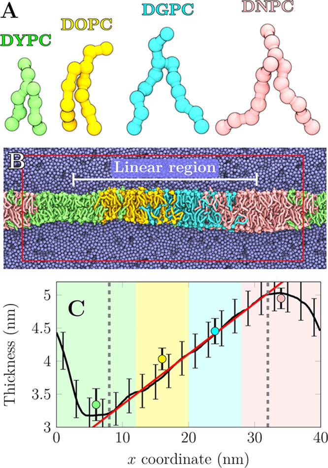 Figure 1
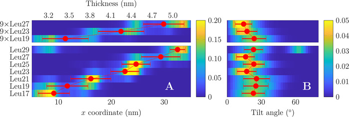 Figure 2
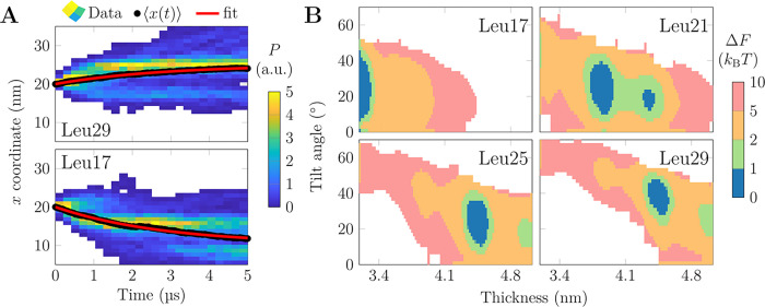 Figure 3
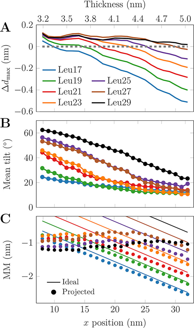 Figure 4
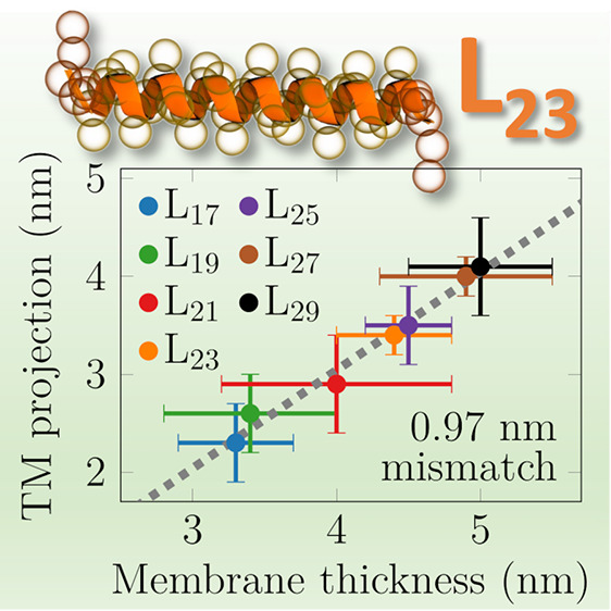 Figure 5
- —Luonnontieteiden ja Tekniikan Tutkimuksen Toimikunta10.13039/501100005877
Peer Reviews
No public reviews on file for this paper yet. If you reviewed it on a platform where reviews are public (OpenReview, ICLR, NeurIPS, ICML), you can paste yours below so the community can read it here.
Videos
No videos yet. Explain this paper in a talk, walkthrough, or lecture? Add one.
Taxonomy
TopicsLipid Membrane Structure and Behavior · Nanopore and Nanochannel Transport Studies · RNA Interference and Gene Delivery
Cellular membranes display varied lipid compositions and hence physicochemical properties supporting the processes taking place in and on them.^1^ The membranes of the endoplasmic reticulum, the Golgi apparatus, and the plasma membrane differ in their thicknesses,^2^ suggesting that hydrophobic mismatch (MM) controls the sorting and targeting of transmembrane (TM) peptides and proteins along the secretory pathway. MM is the difference in the hydrophobic extents of the transmembrane domain (TMD) and the host membrane^3,4^ which—if unaccounted for—leads to an energetic penalty. Apart from organelle-level sorting, MM also affects protein function, conformation, stability, orientation, oligomerization, and dynamics.^5−8^ Moreover, the plasma membrane^9^ and organelle membranes^10^ are heterogeneous with different local lipid pools manifested in different membrane properties—including thickness—leading to lateral sorting of proteins.^11^ Sometimes, MM cannot be eliminated by lateral sorting, but instead either the membrane, the protein, or both adapt to eliminate the mistmatch. For multipass TMDs, this is not straightforward without conformational changes. However, single-pass TMDs can cope with mismatch by multiple mechanisms.
In the case of positive MM (TMD longer than membrane thickness), the TMD can either tilt or bend,^5,12−15^ and the membrane can also respond by locally adjusting its thickness.^16,17^ The latter mechanism is intriguing, since it can drive TMD aggregation by lowering the total energetic penalty associated with membrane deformation.^14^ In the case of negative MM, the membrane can bulge, possibly coupled with TMD aggregation.^13,16,17^ Alternatively, water can penetrate the headgroup region to hydrate charged protein residues.^13^ In extreme MM scenarios, the TMDs might alternate their anchoring between membrane leaflets, or even abandon their TM orientation.^15,16^ Of exceptional interest are the related energetics, as it maps the TMD and membrane properties to the preferred response. For example, a single-pass TMD with MM > 0 might partition to a thicker domain or stay put and increase its tilt. Both actions would at least partially eliminate MM, but which one is favored? Or are both active within the limits of the thermal energy? Surprisingly, these questions have escaped earlier computational and experimental examinations, likely due to methodological challenges.
Here, we tackle these questions using coarse-grained (CG) molecular dynamics (MD) simulations. We set up a lipid membrane containing a thickness gradient by merging four single-component membranes made of different phosphatidylcholine (PC) lipids with increasing acyl chain lengths: DYPC (3 coarse-grained beads per chain), DOPC (4 beads), DGPC (5 beads), and DNPC (6 beads), corresponding to 12–24 carbon atoms per chain in the Martini 2 model^18,19^ (Figure 1A). The thicknesses of corresponding single-lipid membranes—ranging from 3.34 to 4.95 nm—are indicated by markers in Figure 1C. To generate a constant thickness profile along the x axis, the different lipid types are maintained at certain x-coordinate intervals using flat-bottom potentials (Figure 1B). A smooth profile in the x coordinate range of 8–32 nm is generated with an optimized amount of lipid type overlap (Figure 1C; see also methods and Figure S2 in the Supporting Information (SI)), resulting in a maximum thickness difference of ≈1.85 nm.
Our approach extends the toolkit available for the study of hydrophobic mismatch and overcomes some limitations of earlier works. Our setup contains a smooth thickness gradient instead of a two-phase membrane arrangement.^20,21^ Moreover, the use of different lipid types to generate the gradient in our approach allows membranes to be in their native, tensionless state, hence realistic lateral lipid densities canbe studied.^22^ A final remark considers the ease-of-use; unlike the setup in ref (22), a standard GROMACS version is suitable to run the simulations in our approach.
Due to periodic boundary conditions, there is an abrupt thickness jump at the edge of the simulation box, but analyses here focus on the linear region. The shape of the flat-bottom potentials is provided in Figure S1C, whereas Figure S1E shows the resulting spatial distribution of the different lipid types. The local area per lipid values, shown in Figure S1D, are in reasonable agreement with values from single-component membrane simulations. Details of system setup and simulations are available in the SI.
We first studied whether the thickness gradient would sort lipids, as highlighted by similar earlier works.^20,22^ To this end, we released the flat-bottom restraints for 5 lipids of each type in both leaflets, and allowed them to freely sample the membrane during a 50 μs simulation. Curiously, the sorting tendency turned out to be minor with free energy differences of ≈1 kBT (Figure S1F), yet the analysis also revealed a small repulsive bias of similar magnitude (≈1 kBT) at the mixed lipid regions despite our attempts to optimize the overlap of the lipid patches (Figure S2).
We next embedded polyleucines (Leu17–Leu29) whose hydrophobic thicknesses lTM ranged from 2.55 to 4.35 nm to this membrane (Table 1). Polyleucines have demonstrated tolerance for large MM and maintain their TM orientation.^23^ The peptides were capped by two lysines at each end to anchor them to the membrane–water interfaces.^24^ See SI for further details.
We assessed whether the thickness gradient sorts the peptides, or whether alternative mechanisms—such as peptide tilt or membrane deformation—dominate. This is made possible by the thickness gradient, which—unlike biphasic setups^20,21^—will induce a position-dependent lateral force on the peptides. The secondary structure of the peptides is fixed in the Martini 2.2 model,^18,19^ disallowing stretching, bending, or helix breaking, yet these are expected not to be important.^15,16^ We performed 3 sets of 100 μs-long unbiased simulations with varying initial positions of the peptides with a flat-bottom potential maintaining them within the linear regime of the thickness gradient (x = 5–35 nm). As the peptides diffuse at rates of D ≈ 0.015–0.027 nm^2^/ns, they cover δx = 100 nm in the simulation time of Δ = 100 μs, allowing them to spontaneously find their preferred membrane environment, while (large) MM likely renders this search even faster. Rapid sorting indeed occurs as evidenced by the time traces of the peptide positions (Figure S3 and movie at DOI: 10.6084/m9.figshare.24105606). We also repeated this calculation with 9 copies of Leu19, Leu23, or Leu27 present in the membrane and initially placed along the thickness gradient at constant intervals (Figure S4 and DOI: 10.6084/m9.figshare.24105606). The lateral peptide density profiles and tilt angle distributions are shown as colormaps in Figure 2. Red markers show mean ± standard deviation of the position of the peptide center of mass (and hence preferred thickness deq, defined as interleaflet phosphate distance) or tilt angle (θ_eq_), which are also listed in Table 1. In one replica, Leu29 never left its initial position in the thin membrane (Figure S3) and was hence omitted from the calculation of deq and θ_eq_.
The deq values are 0.6–0.9 nm larger than the hydrophobic peptide lengths (lTM). Despite lateral sorting, the peptides still demonstrate a significant tilt θ_eq_ of 22 ± 4° on average without a systematic dependence on peptide length (Figure 2B). Curiously, Leu23 and Leu27 tilt somewhat less (≈17°) than other peptides (≈24°), and this is reproduced in the multipeptide system. Tilting leads to the projected peptide length lproj along the z axis (normal to the membrane) being ≈0.3 nm shorter than lTM. Thus, the realized MM value (lmis = deq – lproj) is consistently 1.0 ± 0.1 nm for all studied peptides, whereas an extrapolation to zero length provides a mismatch value of 0.8 nm (see TOC graphic). Concluding, spontaneous sorting takes place on the simulation time scale, while tilting also contributes to MM elimination.
To obtain physical insight into the sorting, we placed a single Leu17 or Leu29 at the center of the thickness gradient and performed 100 independent unbiased simulations (Figure 3A). We hypothesized that the velocity along the gradient (x) is linearly proportional to the distance of the peptide from its equilibrium location, , in other words proportional to MM. We solve for x = x0 – A(1 – exp(−k/τ)), which fits the data in Figure 3A remarkably well (R^2^ > 0.99). This validates that v ∼ MM, which in viscous overdamped media where F ∼ v also indicates that F ∼ MM, i.e., a harmonic force. Curiously, both Leu17 (here MM < 0) and Leu29 (MM > 0) were found to relax toward xeq with a time constant of 2.8 μs, suggesting that the force due to MM is symmetric around 0. Our peptides mainly respond to MM by tilting, yet membrane thickness deformation due to larger proteins was found to have a similar dependence on mismatch.^25^
The peptides occasionally sample different thicknesses and tilt angles during the unbiased simulations (Figure S3), indicating that the underlying free energy minima are shallow. To verify this, we performed umbrella sampling (US) simulations to extract potentials of mean force (PMFs) for the peptide position and hence as a function of local thickness (ΔF(d)). We combined this PMF with the free energy profile of peptide tilt angle obtained for each US window as ΔF(θ)tilt = −kBT ln(P(θ)/P(θ_0_)) with kBT the thermal energy and θ_0_ the most likely tilt angle in that US window (ΔF(θ_0_) = 0). The sum ΔF(d, θ) ≈ ΔF(d) + ΔF(θ) was used to approximate the 2D free energy surface, and it provides both the minima as well as the thermally accessible ranges of thickness and tilt angle for each peptide (selected ones in Figure 3B, the rest in Figure S5). All profiles demonstrate minima which cover 0.1–0.3 nm in thickness (dkT) and 14–41° in tilt angle (θ_kT_) within ΔF = kBT from the global minimum (ranges listed in Table 1; for peptides with two minima, values for both are reported). The profiles also reveal that doubling the threshold to 2 × kBT barely increases the accessible ranges, whereas within 5 × kBT, the peptides can already sample a thickness range > 1 nm. The accessible tilt angle range is less sensitive. Finally, within the 10 × kBT threshold, the peptides sample the entire available thickness range. The profiles demonstrate the expected trend; the thinner the membrane, the more tilted the peptides.
The entropic contribution leading to isotropic (polar) tilt angle θ dominates,^26^ yet the thickness gradient could also induce directional tilt. We applied the one-sample Kolmogorov–Smirnov test to estimate the p values for the hypothesis that the azimuth angle (φ) of the peptide differed from uniform distribution. The probability distribution of the p values in Figure S6 demonstrates that in some cases the distribution deviates from the uniform one. However, these small p values seem to be distributed randomly among the studied peptides and among membrane thicknesses, indicating that there is no systematic tilt due to the gradient or the overlap regions inherent in our setup.
While the energetics of sorting by MM has not been previously studied, Kim and Im extracted the thermally accessible tilt angle ranges for WALP23/WALP27 peptides in POPC and DMPC membranes.^12^ WALP23/WALP27 have lTM values similar to our Leu17/Leu21 peptides. From separate simulations, we determined the thickness of single-component POPC and DMPC membranes and identified the US windows with similar thicknesses.In the window matching the thickness of POPC, the Leu17/Leu21 sample tilt angles of 5–29°/10–36°, whereas the values for WALP23/WALP27 in POPC were similar at 7–26°/14–46°.^12^ For DMPC-like thickness, Leu17/Leu21 showed tilts of 6–34°/18–47°, in reasonable agreement with WALP23/WALP27 in DMPC with 14–39°/32–51°.^12^ The small differences likely arise from different peptide sequences, especially the residues anchoring them to the membrane–water interfaces.
The US windows provide a systematic set of different (fixed) MM conditions. We used these windows to study how the host membrane responds to the presence of the peptides of various lengths. We extracted the 2-dimensional thickness maps around for each peptide and for each US window. The thickness perturbations as a function of US window (peptide location) and membrane location are shown in Figure S7. These maps demonstrate that the perturbed region spans a few nanometers around the peptide. We also extracted the values on the diagonal of these maps, i.e., the thickness perturbation values at the peptide position, which are shown in Figure 4A. These values clearly indicate that even the longest peptides have a meager (≈0.1 nm) thickening effect, whereas the shortest peptides render the thick membrane regions thinner by ≈0.5 nm). This asymmetry is perhaps not surprising, as the thickening is limited by the length of the acyl chains in their extended conformation, whereas thinning can occur to a larger extent via acyl chain tilt, disordering, or even their interdigitation. This thinning contributes to MM estimates, but due to its complexity, we have omitted it from our other analyses. We next analyzed how the peptides respond to MM via tilting. Figure 4B demonstrates tilt angles larger than 60° in the case of significant positive MM. With thicker membranes, the tilt angles decrease monotonously until saturation at ≈10°. Our membrane does not contain a thick enough region to observe this saturation for the three longest peptides. We used these tilt values to calculate the projected peptide lengths (lproj) along the z axis (membrane normal), as for the unbiased simulations (Table 1). We then estimated the real MM defined as lmis = deq – lproj (Figure 4C). Ideal MM (assuming peptide orientation along membrane normal) is also shown. In the case of positive ideal MM, peptide tilting leads to a fairly constant projected MM of −1 nm, in line with our unbiased simulations (Table 1). There is no obvious peptide length dependence. In the thicker membrane with ideal MM < −1 nm, the peptides can no longer tilt but rather maintain a constant tilt (Figure 4B), and the projected MM follows the ideal behavior. Curiously, the magnitude of lproj for each peptide is smallest right before it starts to follow the ideal MM scenario, i.e., at the least tilted conformation. This suggests that in too thin membranes, the peptides overcompensate for the MM by tilting.
Concluding, we have presented a novel simulation approach to study phenomena affected by membrane thickness. Using a combination of different lipid species and flat-bottom restraints, a thickness gradient is maintained along one axis of the simulation box. As the first example, we have focused on single-span peptides, which serve as model systems for the TMDs of physiologically important receptor tyrosine kinases. Our results demonstrate that peptides of different lengths are spontaneously sorted over distances of dozens of nanometers on the microsecond time scale. This indicates that our setup can be efficiently used to study the sorting of lipids, peptides, proteins, and other membrane-embedded objects. It can also be easily adapted to the study of larger membrane-spanning objects. However, for major protein complexes, the membrane dimensions might have to be extended, requiring the calibration of the overlap of the neighboring phases, i.e., the adjustment of the widths of the flat-bottom potentials.
Moreover, with the x coordinate one-to-one mapped to thickness, conformational changes of proteins induced by the latter can be readily studied. Free energy profiles of thickness-dependent properties—such as sorting or conformation—can be extracted using a simple reaction coordinate. We facilitate these and other yet unconsidered applications by providing all the simulation inputs and outputs in the Zenodo repository at DOIs: 10.5281/zenodo.10887673 and 10.5281/zenodo.10840054. Here, we used the CG Martini^18,19^ model, yet the future extension to atomistic resolution is straightforward. Morever, the CG approach has limited resolution, and although the smoothness of our thickness gradient is optimized (Figure S2), there is still a small yet detectable bias of both the lipids (Figure S1F) and the peptides (Figure 3) toward the single-component membrane regions, rendering our approach only semiquantitative. Other proposed approaches for the study of lipid sorting by mismatch seem to also suffer from boundary effects.^20−22^ In our setup, this is potentially an entropic effect, as a peptide in the overlap region decreases the total amount of lipid mixing permitted within the flat-bottom restraints. This is likely overcome with the additional resolution and hence smoother profiles of atomistic models.
The reference list from the paper itself. Each links out to its DOI / PubMed record.
- 1Van Meer G.; Voelker D. R.; Feigenson G. W. Membrane lipids: Where They Are and How They Behave. Nat. Rev. Mol. Cell Biol. 2008, 9, 112–124. 10.1038/nrm 2330.18216768 PMC 2642958 · doi ↗ · pubmed ↗
- 2Mitra K.; Ubarretxena-Belandia I.; Taguchi T.; Warren G.; Engelman D. M. Modulation of the Bilayer Thickness of Exocytic Pathway Membranes by Membrane Proteins Rather Than Cholesterol. Proc. Natl. Acad. Sci. U.S.A. 2004, 101, 4083–4088. 10.1073/pnas.0307332101.15016920 PMC 384699 · doi ↗ · pubmed ↗
- 3Schmidt U.; Weiss M. Hydrophobic Mismatch-Induced Clustering as a Primer for Protein Sorting in the Secretory Pathway. Biophys. Chem. 2010, 151, 34–38. 10.1016/j.bpc.2010.04.009.20537786 · doi ↗ · pubmed ↗
- 4Grau B.; Javanainen M.; García-Murria M. J.; Kulig W.; Vattulainen I.; Mingarro I.; Martínez-Gil L. The Role of Hydrophobic Matching on Transmembrane Helix Packing in Cells. Cell Stress 2017, 1, 9010.15698/cst 2017.11.111.31225439 PMC 6551820 · doi ↗ · pubmed ↗
- 5Killian J. A. Hydrophobic Mismatch Between Proteins and Lipids in Membranes. Biochim. Biophys. Acta 1998, 1376, 401–416. 10.1016/S 0304-4157(98)00017-3.9805000 · doi ↗ · pubmed ↗
- 6Jensen M. Ø.; Mouritsen O. G. Lipids do Influence Protein Function—The Hydrophobic Matching Hypothesis Revisited. Biochim. Biophys. Acta 2004, 1666, 205–226. 10.1016/j.bbamem.2004.06.009.15519316 · doi ↗ · pubmed ↗
- 7Ramadurai S.; Duurkens R.; Krasnikov V. V.; Poolman B. Lateral Diffusion of Membrane Proteins: Consequences of Hydrophobic Mismatch and Lipid Composition. Biophys. J. 2010, 99, 1482–1489. 10.1016/j.bpj.2010.06.036.20816060 PMC 2931744 · doi ↗ · pubmed ↗
- 8Castillo N.; Monticelli L.; Barnoud J.; Tieleman D. P. Free Energy of WALP 23 Dimer Association in DMPC, DPPC, and DOPC Bilayers. Chem. Phys. Lipids 2013, 169, 95–105. 10.1016/j.chemphyslip.2013.02.001.23415670 · doi ↗ · pubmed ↗