Multimodal imaging assessment for feasibility of endovascular reconstruction in cases of inferior vena cava atresia
Adam Kisling, Sean Basile, Adelle Dagher, Jonathan Sexton, Eric Twerdahl

TL;DR
This paper discusses using advanced imaging techniques to assess the feasibility of a specific endovascular procedure for a rare vein condition.
Contribution
The study highlights the role of multimodal imaging in planning endovascular reconstruction for inferior vena cava atresia.
Findings
Multimodal imaging is essential for evaluating the complex anatomy in inferior vena cava atresia.
CT and venography help determine the suitability of endovascular intervention.
These imaging techniques aid in detailed procedural planning for affected patients.
Abstract
Inferior vena cava atresia is a rare condition with highly variable anatomy due to the complexity of caval embryology. When endovascular venovenous reconstruction is considered for severe persistent sequelae, multimodality imaging with CT and invasive venography is used to determine the appropriateness of intervention and for procedural planning.
Genes, proteins, chemicals, diseases, species, mutations and cell lines named across the full text — each resolved to its canonical identifier and authoritative record.
Click any figure to enlarge with its caption.
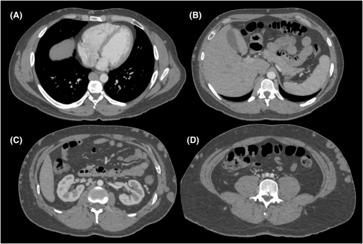 Figure 1
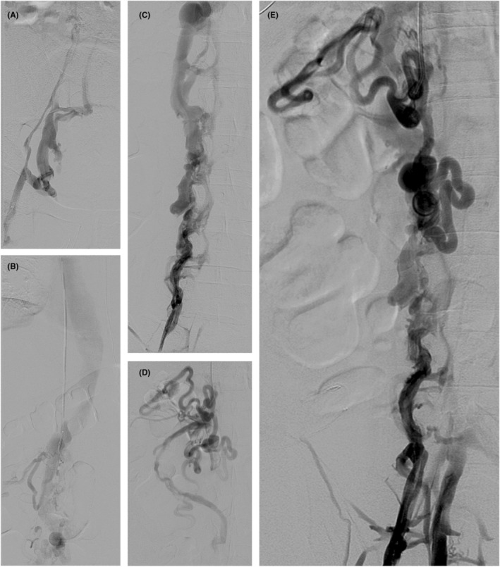 Figure 2
Peer Reviews
No public reviews on file for this paper yet. If you reviewed it on a platform where reviews are public (OpenReview, ICLR, NeurIPS, ICML), you can paste yours below so the community can read it here.
Videos
No videos yet. Explain this paper in a talk, walkthrough, or lecture? Add one.
Taxonomy
TopicsVascular anomalies and interventions · Central Venous Catheters and Hemodialysis · Venous Thromboembolism Diagnosis and Management