A Pathophysiological Approach to Spontaneous Orbital Meningoceles: Case Report and Systematic Review
Piergiorgio Gaudioso, Elia Biancoli, Veronica Battistuzzi, Stefano Concheri, Tommaso Saccardo, Sebastiano Franchella, Giacomo Contro, Stefano Taboni, Elisabetta Zanoletti, Francesco Causin, Lorena Nico, Joseph Domenico Gabrieli, Roberto Maroldi, Piero Nicolai, Marco Ferrari

TL;DR
This paper reports a rare case of an orbital meningocele in a child and reviews existing literature to emphasize the importance of understanding its pathophysiology for treatment.
Contribution
The study presents a new clinical case and provides a systematic review of spontaneous orbital meningoceles, highlighting treatment approaches and pathophysiological insights.
Findings
A 6-year-old patient with orbital meningocele showed clinical improvement after endovascular thrombectomy and stenting.
The systematic review found that surgery is the most common treatment for spontaneous orbital meningoceles.
The paper emphasizes the need for further data collection due to the limited number of reported cases.
Abstract
Background: Spontaneous orbital cephaloceles are a rare condition. The purpose of this study is to provide a description of a clinical case and to carry out a systematic literature review. Methods: A systematic review of the English literature published on the Pubmed, Scopus, and Web of Science databases was conducted, according to the PRISMA recommendations. Results: A 6-year-old patient was admitted for right otomastoiditis and thrombosis of the sigmoid and transverse sinuses, as well as the proximal portion of the internal jugular vein. Radiological examinations revealed a left orbital mass (22 × 14 mm) compatible with asymptomatic orbital meningocele (MC) herniated from the superior orbital fissure (SOF). The child underwent a right mastoidectomy. After the development of symptoms and signs of intracranial hypertension (ICH), endovascular thrombectomy and transverse sinus stenting…
Genes, proteins, chemicals, diseases, species, mutations and cell lines named across the full text — each resolved to its canonical identifier and authoritative record.
Click any figure to enlarge with its caption.
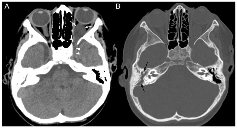 Figure 1
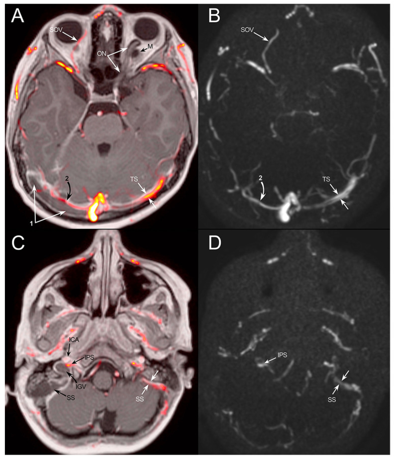 Figure 2
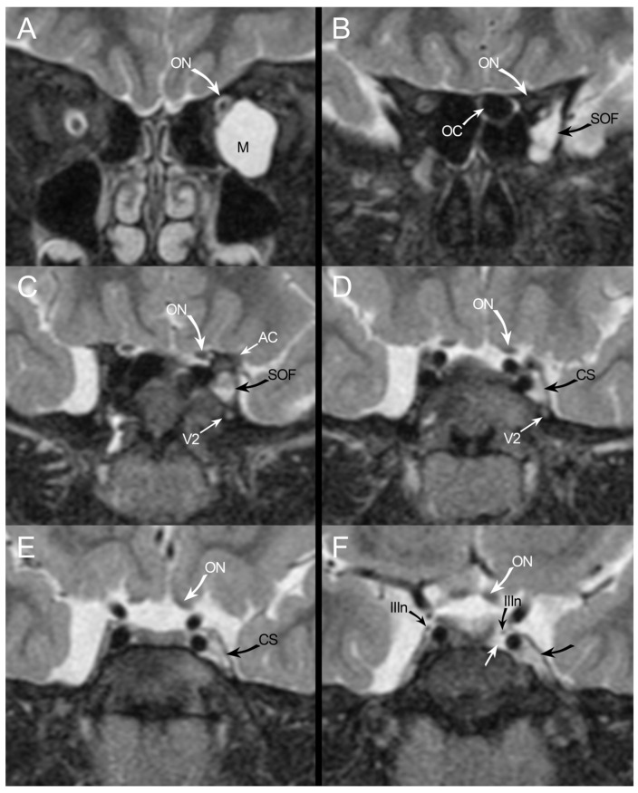 Figure 3
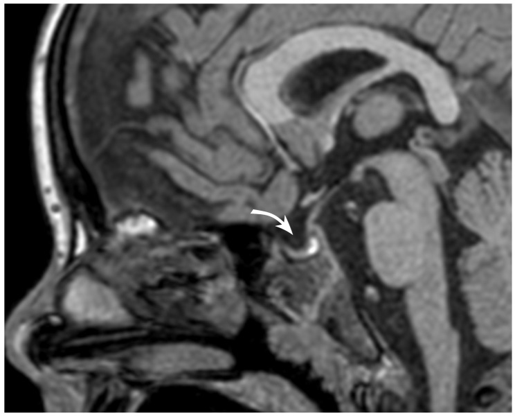 Figure 4
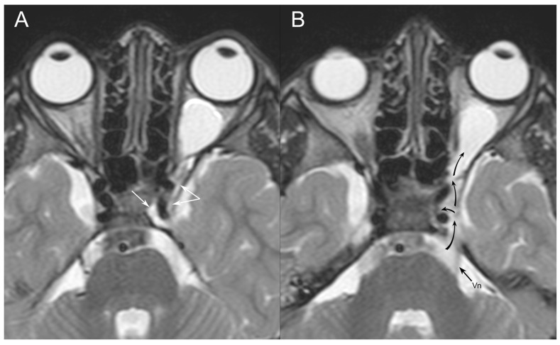 Figure 5
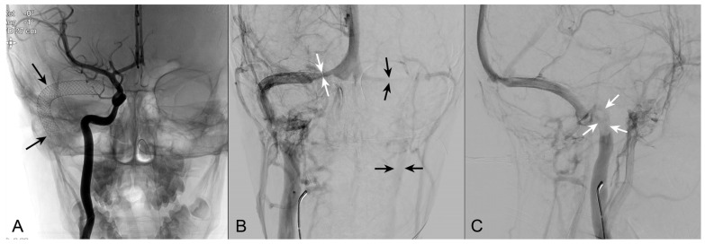 Figure 6
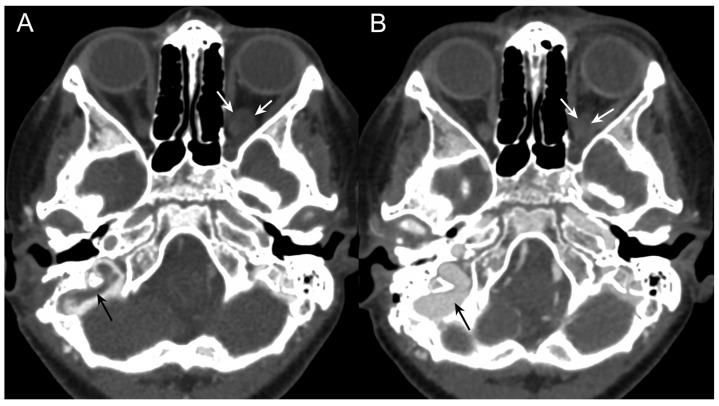 Figure 7
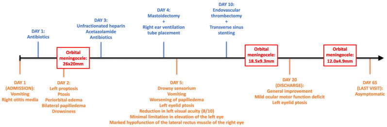 Figure 8
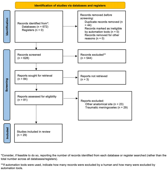 Figure 9
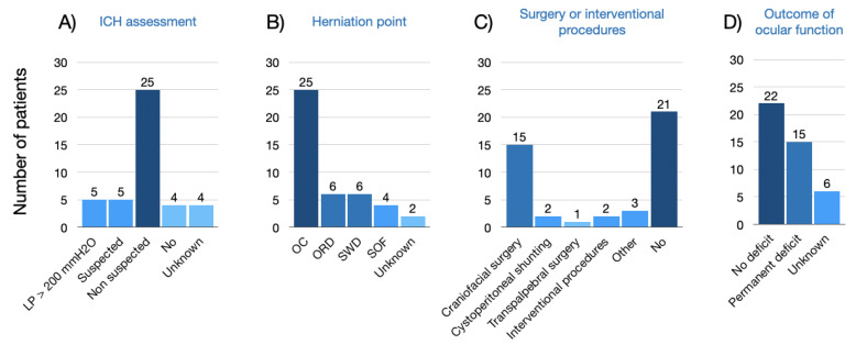 Figure 10
Peer Reviews
No public reviews on file for this paper yet. If you reviewed it on a platform where reviews are public (OpenReview, ICLR, NeurIPS, ICML), you can paste yours below so the community can read it here.
Videos
No videos yet. Explain this paper in a talk, walkthrough, or lecture? Add one.
Taxonomy
TopicsCerebral Venous Sinus Thrombosis · Head and Neck Surgical Oncology · Meningioma and schwannoma management