Improving the Generalizability of Deep Learning for T2-Lesion Segmentation of Gliomas in the Post-Treatment Setting
Jacob Ellison, Francesco Caliva, Pablo Damasceno, Tracy L. Luks, Marisa LaFontaine, Julia Cluceru, Anil Kemisetti, Yan Li, Annette M. Molinaro, Valentina Pedoia, Javier E. Villanueva-Meyer, Janine M. Lupo

TL;DR
This paper improves deep learning models for tracking brain tumor changes after treatment by using mixed data and new techniques to better segment T2-lesions in MRI scans.
Contribution
The study introduces data mixing, transfer learning, and spatial regularization to enhance T2-lesion segmentation in post-treatment glioma MRI scans.
Findings
Including 26% treated patients in training improved performance by 13.9%.
Fine-tuning with treated glioma data improved sensitivity by 2.5% compared to data mixing.
Spatial regularization improved performance metrics like HD, Dice, and sensitivity when used with transfer learning.
Abstract
Although fully automated volumetric approaches for monitoring brain tumor response have many advantages, most available deep learning models are optimized for highly curated, multi-contrast MRI from newly diagnosed gliomas, which are not representative of post-treatment cases in the clinic. Improving segmentation for treated patients is critical to accurately tracking changes in response to therapy. We investigated mixing data from newly diagnosed (n = 208) and treated (n = 221) gliomas in training, applying transfer learning (TL) from pre- to post-treatment imaging domains, and incorporating spatial regularization for T2-lesion segmentation using only T2 FLAIR images as input to improve generalization post-treatment. These approaches were evaluated on 24 patients suspected of progression who had received prior treatment. Including 26% of treated patients in training improved…
Genes, proteins, chemicals, diseases, species, mutations and cell lines named across the full text — each resolved to its canonical identifier and authoritative record.
Click any figure to enlarge with its caption.
 Figure 1
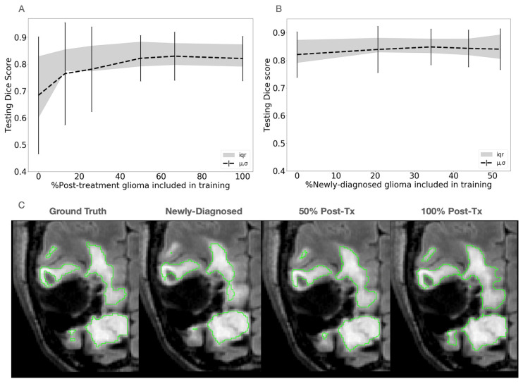 Figure 2
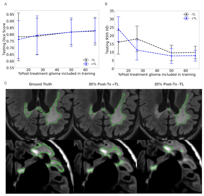 Figure 3
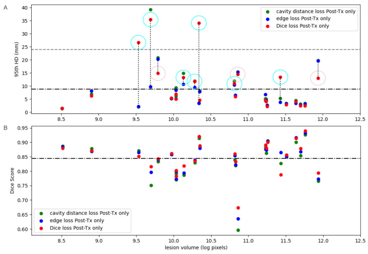 Figure 4
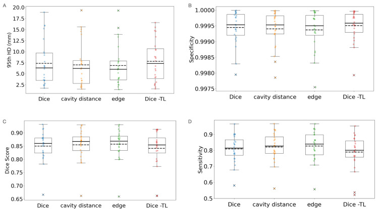 Figure 5
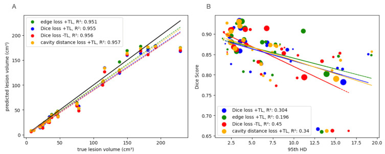 Figure 6
 Figure 7
 Figure 8
Peer Reviews
No public reviews on file for this paper yet. If you reviewed it on a platform where reviews are public (OpenReview, ICLR, NeurIPS, ICML), you can paste yours below so the community can read it here.
Videos
No videos yet. Explain this paper in a talk, walkthrough, or lecture? Add one.
Taxonomy
TopicsBrain Tumor Detection and Classification · Medical Image Segmentation Techniques · Advanced Neural Network Applications