Medial sigmoid depression prevalence and association with a sigmoid notch: cone beam computed tomography and panoramic image study
Ozlem Busra Dogan, Hatice Boyacioglu

TL;DR
This study compares how often a specific anatomical feature appears in two imaging techniques and finds that both methods show similar results.
Contribution
The study demonstrates that panoramic imaging can reliably detect medial sigmoid depression as effectively as CBCT.
Findings
MSD prevalence was 66.7% for CBCT and 58.1% for panoramic imaging.
MSD prevalence did not significantly differ between males and females.
MSD shape and prevalence were not associated with sigmoid notch morphology.
Abstract
This study aims to determine whether and how the data of the medial sigmoid depression (MSD) area via cone beam computed tomography (CBCT) differs from panoramic radiography. This study also aims to evaluate various sigmoid notch types and assess the relationship between sigmoid depression and notch morphology. A total of 129 individuals consisting of 258 sides were evaluated. Chi-Square/Fisher Exact tests were used to assess parameters on a categorical scale between two or more groups. McNemar’s test compared the findings detected on panoramic and CBCT images. MSD was more prevalent in females than males in both techniques, but this difference was not statistically significant. There was no association between the prevalence of MSD and the morphology of the sigmoid notch. The incidence of MSD shape was not significantly different between both imaging modalities. In both panoramic and…
Genes, proteins, chemicals, diseases, species, mutations and cell lines named across the full text — each resolved to its canonical identifier and authoritative record.
Click any figure to enlarge with its caption.
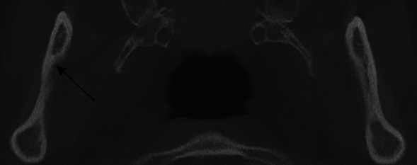 Figure 1
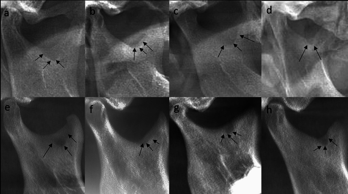 Figure 2
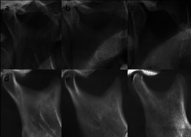 Figure 3
Peer Reviews
No public reviews on file for this paper yet. If you reviewed it on a platform where reviews are public (OpenReview, ICLR, NeurIPS, ICML), you can paste yours below so the community can read it here.
Videos
No videos yet. Explain this paper in a talk, walkthrough, or lecture? Add one.
Taxonomy
TopicsDental Radiography and Imaging · Facial Trauma and Fracture Management · Facial Nerve Paralysis Treatment and Research