A case of endoscopic ultrasonography-guided choledochoduodenostomy for malignant distal bile duct obstruction with upside-down stomach
Takuya Takayanagi, Yusuke Sekino, Kota Ueno, Shota Matsumoto, Noriki Kasuga, Hajime Nagase

Abstract
Genes, proteins, chemicals, diseases, species, mutations and cell lines named across the full text — each resolved to its canonical identifier and authoritative record.
Click any figure to enlarge with its caption.
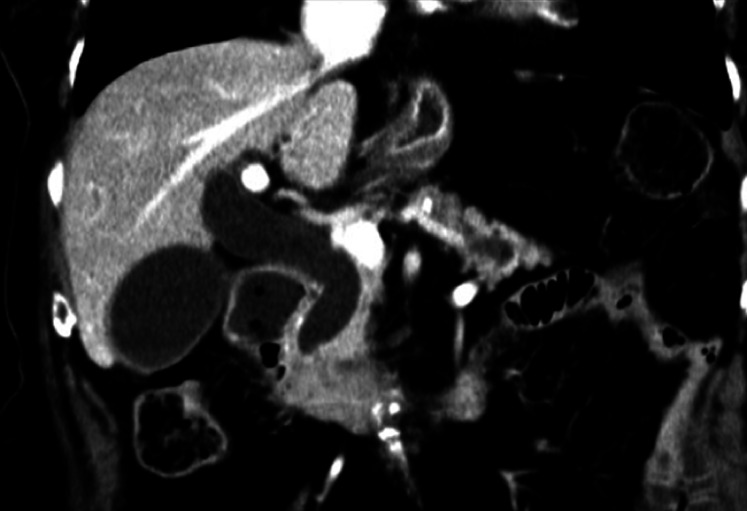 Fig. 1
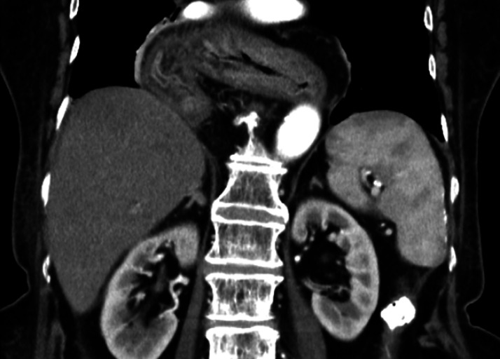 Fig. 2
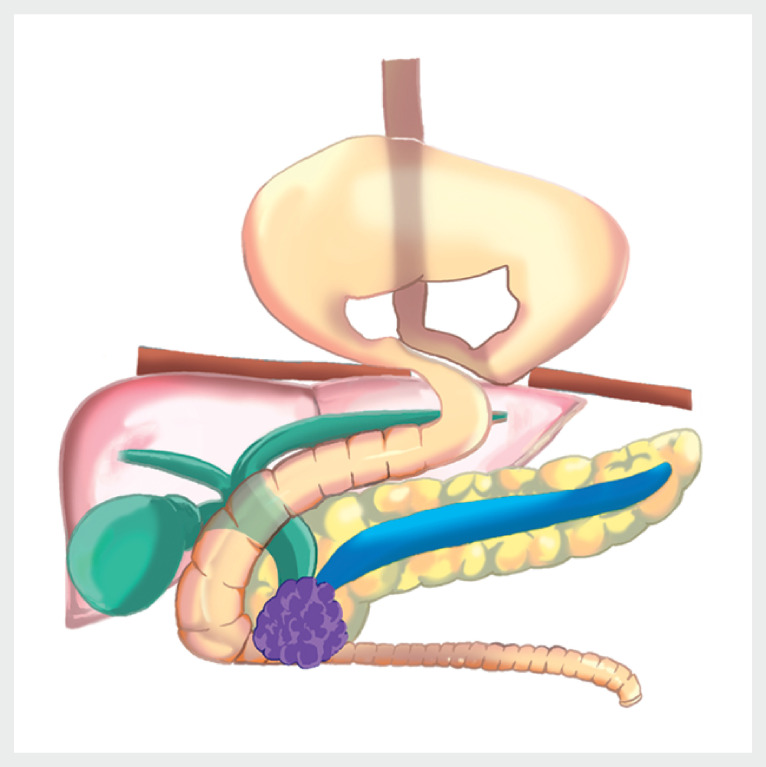 Fig. 3
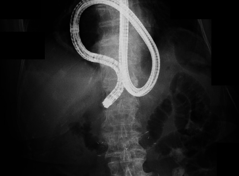 Fig. 4
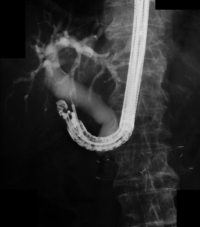 Fig. 5
Peer Reviews
No public reviews on file for this paper yet. If you reviewed it on a platform where reviews are public (OpenReview, ICLR, NeurIPS, ICML), you can paste yours below so the community can read it here.
Videos
No videos yet. Explain this paper in a talk, walkthrough, or lecture? Add one.
Taxonomy
TopicsPediatric Hepatobiliary Diseases and Treatments · Esophageal and GI Pathology · Gastrointestinal disorders and treatments
Endoscopic ultrasonography-guided biliary drainage (EUS-BD) is increasingly being used as a drainage technique in cases where endoscopic retrograde cholangiopancreatography (ERCP) cannot be performed. However, in patients with a massive esophageal hiatal hernia, this poses a risk of mediastinitis due to thoracic puncture. Upside-down stomach, first described in 1926 as type IV diaphragmatic hiatal hernia, is a rare condition in which a large portion of the stomach migrates into the thoracic cavity due to organoaxial rotation 1 . There have been reports of ERCP in patients with upside-down stomach 2 3 but none of EUS-BD. This is the first report of EUS-BD for distal bile duct obstruction in a patient with pancreatic head cancer and upside-down stomach.
An 84-year-old woman presented to our hospital with the chief complaint of weight loss. Computed tomography revealed a 35-mm tumor in the pancreatic head, and obstruction of the distal bile duct and duodenum by the tumor was suspected ( Fig. 1 ). The patient also had an esophageal hiatal hernia of the upside-down stomach type, and almost all of her stomach had prolapsed into the thoracic cavity ( Fig. 2 , Fig. 3 ). We decided to attempt endoscopic ultrasonography-guided choledochoduodenostomy (EUS-CDS) along with duodenal stent placement in the papillary region.
Computed tomography revealed a 35-mm tumor in the pancreatic head, and obstruction of the distal bile duct and duodenum by the tumor was suspected.
The patient also had a esophageal hiatal hernia of the upside-down stomach type, and almost all of her stomach had prolapsed into the thoracic cavity.
Schema of this case.
A forward-viewing scope was advanced to the duodenum, and a stiff-type guidewire was advanced to the jejunum ( Fig. 4 ). The guidewire was left in place, and the endoscope was removed while the loop for the upside-down stomach was released. A convex endoscope (UCT-260; Olympus, Tokyo, Japan) was then advanced over the wire to the esophagogastric junction. The contrast catheter was advanced, and the endoscope was successfully maneuvered around it to the duodenum. Thereafter, EUS-CDS was performed, and a covered self-expanding metal stent (ZEO STENT, 8 × 60 mm; Zeon Medical Inc., Tokyo, Japan) was placed successfully ( Video 1 , Fig. 5 ). This case demonstrates that EUS-BD can be safely performed for malignant distal bile duct obstruction with upside-down stomach if EUS-CDS is used.
A forward-viewing scope was advanced to the duodenum, and a stiff-type guidewire was advanced to the jejunum. The guidewire was left in place, and the endoscope was removed while the upside-down stomach loop was released.
A convex endoscope was successfully maneuvered around it to the duodenum. Thereafter, EUS-CDS was performed, and a covered self-expanding metal stent was placed successfully.
Endoscopic ultrasonography-guided choledochoduodenostomy for malignant distal bile duct obstruction with upside-down stomach.Video 1
Endoscopy_UCTN_Code_TTT_1AS_2AH
The reference list from the paper itself. Each links out to its DOI / PubMed record.
- 1Akerlund A Hernia diafragmatica hiatus oesophagei vom anatomischen und röntgenologischen Gesichtspunkt Acta Radiol 19266322
- 2Itoi T Watanabe H Gotoda T Therapeutic endoscopic retrograde cholangiopancreatography using a large dilating balloon in a patient with upside-down stomach and bile duct stones (with video)J Hepatobiliary Pancreat Sci 20152217717925345391 10.1002/jhbp.172 · doi ↗ · pubmed ↗
- 3Khirfan KA rare cause of difficult endoscopic retrograde cholangiopancreatography Gastroenterology 2020158 e 10e 1110.1053/j.gastro.2019.10.04231738919 · doi ↗ · pubmed ↗