Effective xanthine oxidase inhibitor urate lowering therapy in gout is linked to an emergent serum protein interactome of complement activation and inflammation modulators
Concepcion Sanchez, Anaamika Campeau, Ru Liu-Bryan, Ted R Mikuls, James R O'Dell, David J Gonzalez, Robert Terkeltaub

TL;DR
This study shows that urate-lowering therapy for gout leads to changes in blood proteins linked to reduced inflammation and flare-ups.
Contribution
The study identifies a treatment-emergent serum protein network linked to anti-inflammatory effects of xanthine oxidase inhibitors in gout.
Findings
Serum urate normalized and flares declined after 48 weeks of ULT.
Key proteins like C5, IL-1B, and CXCL8 were part of a treatment-emergent interactome.
Febuxostat inhibited complement activation in cultured macrophages.
Abstract
Urate-lowering treatment (ULT) to target with xanthine oxidase inhibitors (XOIs) paradoxically causes early increase in gouty arthritis flares. Because delayed reduction in flare burden is mechanistically unclear, we tested for ULT inflammation responsiveness markers. Unbiased proteomics analyzed blood samples (baseline, 48 weeks ULT) in two, independent ULT out trial cohorts (n = 19, n = 30). STRING-db and multivariate analyses supplemented determinations of altered proteins via Wilcoxon matched pairs signed rank testing in XOI ULT responders. Mechanistic studies characterized proteomes of cultured XOI-treated murine bone marrow macrophages (BMDMs). At 48 weeks ULT, serum urate normalized in all gout patients, and flares declined, with significantly altered proteins (p < 0.05) in clustering and proteome networks in sera and peripheral blood mononuclear cells. Serum proteome changes…
Genes, proteins, chemicals, diseases, species, mutations and cell lines named across the full text — each resolved to its canonical identifier and authoritative record.
Click any figure to enlarge with its caption.
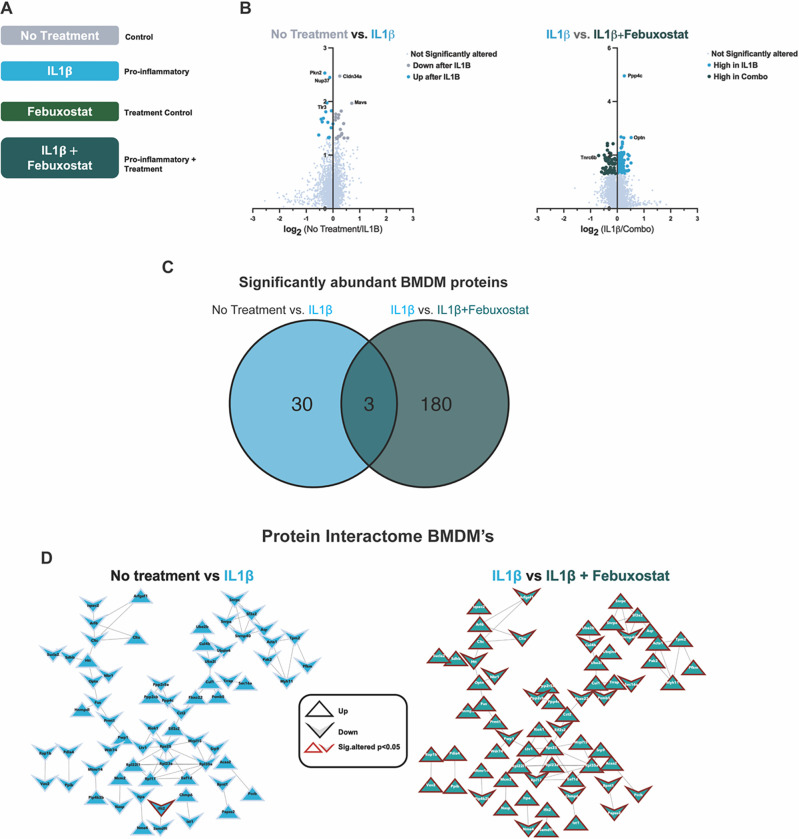 Figure 1
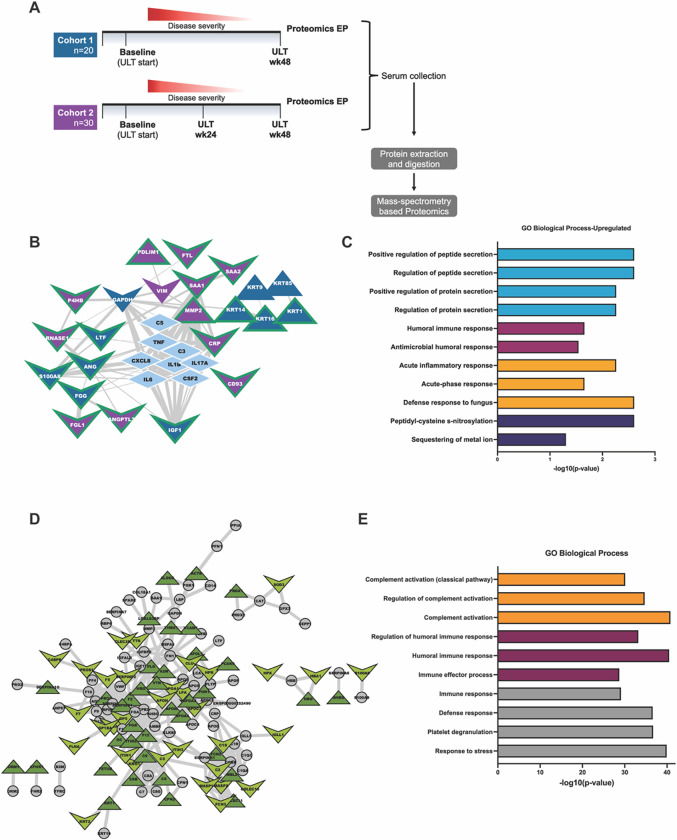 Figure 2
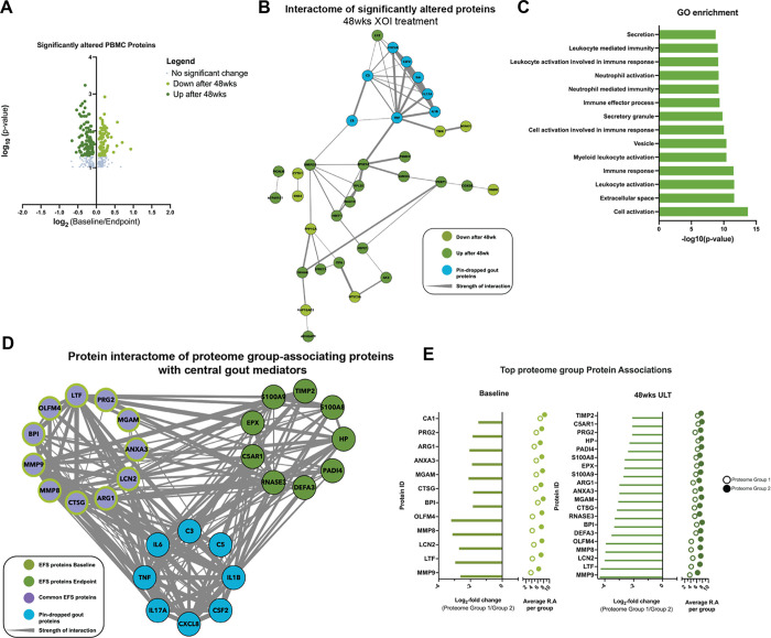 Figure 3
- —NIH/NIAMS
- —VA Research Service
- —UCSD Collaborative Center for Multiplexed Proteomics, and Janssen Pharmaceuticals
- —VA Research Service, NIH
Peer Reviews
No public reviews on file for this paper yet. If you reviewed it on a platform where reviews are public (OpenReview, ICLR, NeurIPS, ICML), you can paste yours below so the community can read it here.
Videos
No videos yet. Explain this paper in a talk, walkthrough, or lecture? Add one.
Taxonomy
TopicsGout, Hyperuricemia, Uric Acid · Inflammasome and immune disorders · Case Reports on Hematomas
Introduction
Gout is characterized by acute arthritis flares that typically are excruciatingly painful and incapacitating^1,2^. Exogenous factors, including joint trauma, certain dietary excesses, and alcohol consumption, can trigger flares^3–5^. Gout flares require treatment with nonsteroidal anti-inflammatory drugs, corticosteroids, and colchicine, which are nonselective, frequently toxic, and interact frequently with other medications ^1,6,7^. Undertreated, gout commonly progresses to more frequent flares, chronic arthritis, and permanent joint damage ^1^. Gout also is linked to prevalent comorbidities mediated by low-grade inflammation (eg, obesity, type 2 diabetes, atherosclerosis, chronic kidney disease)^1,8^.
Pharmacologic treatment of hyperuricemia, most commonly prescribed using XOI drugs (principally allopurinol or febuxostat), is central to gout management^6,7^. However, effective XOI urate-lowering treatment (ULT) to target also paradoxically induces an elevated gout flare burden early in treatment ^6,7,9^. Remodeling of articular monosodium urate (MSU) crystal deposits and consequent release of free crystals are held partly responsible^10–12^. Notably, changes in a subset of CD14 positive monocytes, overactivation of CD8 + T cells, and upregulate arachidonate metabolism also have been implicated perpetuating systemic gouty inflammation after ULT initiation^13^.
MSU crystals stimulate gouty inflammation in large part by activating monocytes and macrophages, promoting NLRP3 inflammasome-mediated IL-1b release, and neutrophil influx and activation that amplify the inflammatory cascade^1,14^. C5 cleavage on the MSU crystal surface, and consequent C5b-9 complement membrane attack complex (MAC) assembly and membrane pore-forming activity play a major role in the model gouty arthritis inflammatory process^15,16^.
Recent clinical trials have demonstrated that effective XOI urate-lowering treatment (ULT) to target eventually reduces gout flare burden and synovitis between 1–2 years therapy^17–19^. Importantly, flares decrease in this time frame. despite total resolution of urate crystal deposits being far slower and particularly difficult to achieve^10^, and despite continuing systemic inflammation even in the periods between flares and in clinical remission^13^. In clinical practice, this situation is associated with lack of clarity on how long anti-inflammatory gout flare prophylaxis, typically using low dose colchicine, is necessary after initiating ULT and achieving the serum urate target^9^.
Significantly, XOI drugs exert anti-inflammatory effects in monocytes and some other cells, including by antioxidant and urate-lowering effects^20–24^. For example, XOI drugs inhibit NLRP3 inflammasome activation, IL-1b release, and chemokine expression in cultured monocyte/macrophage lineage cells^20,21^. In vivo, XOI drugs limit mouse models of atherosclerosis, nonalcoholic steatohepatosis, and certain other diseases involving low-grade chronic inflammation and oxidative stress processes^20–24^. Hence, we conducted a seminal study to test the hypothesis that sustained, effective ULT remodels inflammatory networks in gout by 48 weeks therapy, that XOI could contribute to this effect, and that this could be detectable using unbiased proteomics.
The data revealed the ability of proteomics to detect anti-inflammatory changes in cultured XOI-treated macrophages, and in response to sustained, effective XOI-based ULT in gout patient sera and PBMCs. Our results provide unbiased evidence that sustained treat to target ULT in gout affects complement activation and other inflammatory pathways, and that XOI inhibition may contribute to remodeling of pathways that regulate gouty inflammation.
Methods
Subjects
As previously reported in detail^25^, Cohort 1 and Cohort 2 human subjects were studied under informed consent, and with local IRB approval (at the Jennifer Moreno San Diego Veterans Affairs Medical Center, and at the University of Nebraska Medical Center, respectively). All experiments were performed in accordance with relevant guidelines and regulations. Human subjects samples and clinical and clinical laboratory data were collected specifically in prospective study ancillary to the national, multi-site comparative effectiveness ULT trial VA CSP594 STOP GOUT, whose protocol and CONSORT statement were previously published.^19^ In that trial, gout patients were randomized to a treat to urate target ULT regimen using allopurinol or the more selective XOI febuxostat. Unless contraindicated, colchicine was prescribed as the primary anti-inflammatory gout flare prophylaxis, with colchicine routinely stopped at 6 months ULT. Twenty consecutive patients meeting the 2015 ACR/EULAR gout classification criteria^26^, and with current hyperuricemia, were recruited from the Rheumatology Outpatient Clinic at the San Diego site^25^. Once again^25^, the gout validation cohort (Cohort 2, n = 30)) was from the University of Nebraska Medical Center, in Omaha, NE research site, under informed consent and with local IRB approval. Subjects with active flare, or CRP elevated over 2 mg/L at study onset and endpoints were not excluded from analyses. We previously characterized Cohort 1 gout patient metabolomic profiles at time zero and 12 and 24 weeks of treat to target ULT, done in a blinded way for the XOI used, and following the trial protocol^25^.
Proteomics:
Sera were obtained from both cohorts, with peripheral blood mononuclear cells (PBMCs) also prepared from Cohort 1 samples. All subjects were clinically assessed by study physicians for palpable tophaceous disease and presence of active flare or quiescent arthritis, with co-morbidities and current medications also recorded.
For serum collection, research personnel collected non-fasting blood samples into 10 ml BD Vacutainer Blood Collection Tubes containing spray-coated silica and a polymer gel to facilitate serum separation. Following 30 min incubation at room temperature, tubes were centrifuged for 10 min at 2000×g and sera were transferred into 1.7ml tubes and immediately frozen and stored at − 80°C until analyses were performed.
For PBMC preparation, non-fasting blood samples collected into 10 ml BD Vacutainer K2 EDTA Plus Blood Collection Tubes were transferred to a conical tube containing equal volume of PBS (~ total 20 ml). The samples were then layered over Sigma Histopaque^®^-1077 (20 mL) in 50 mL conical tubes at room temperature, followed by centrifugation at 400×g in a swinging bucket centrifuge for 30 minutes at room temperature with no brake. The white cellular layer containing PBMCs at the interface between the plasma and density gradient was collected and washed in PBS by dilution and centrifugation for 10 minutes at 250×g. PBMC pellets were immediately frozen and stored at − 80°C until analyzed.
Mass Spectrometry Proteomics:
Sample preparation for proteomic analyses of BMDMs and patient sera was done as we previously described in extensive detail^27^, with slight modification to the sample digestion protocol, which used 10μg trypsin in 50mM TEAB at 47°C for 3 hours. After protein extraction and trypsin digest, 50ug aliquots of samples were reserved for TMT pro-labeling^27^. Bridge channels for downstream data analysis of serum samples, were prepped by combining 5μg of all samples; 50μg aliquots of our bridge sample were then prepared for each TMT-plex (5 total).
Mass spectrometry data acquisition
Serum and BMDM proteomic data were acquired as described in detail^27^. In brief, serum and BMDM proteomic data were acquired through an Thermo Orbitrap Fusion equipped with a Thermoeasy nLC 1000. For Mass spectrometry data search, raw mass spectrometry files were searched using Proteome Discoverer 2.5.0.400. The SEQUEST algorithm was used for spectral matches of raw data with in silico generated protein databases. Serum samples were searched against the UniProt Homo sapiens proteome (05–06-2023) and BMDM samples were searched against the Mus musculus proteome (05–06-23).
Mass Spectrometry Metabolomics
Sample preparation of patient sera for metabolomics were essentially as previously described^27^. In brief, for data Analysis, metabolite features were first normalized to the intensity of value of the internal standard, sulfamethazine, in each sample and then multiplied by 1E6. Missing values (with peak intensities of 0) in metabolite features were set to NA. Then, features with more than 20% missing values per group (timepoint) were removed from analysis. Missing values in remaining features were imputed using K-Nearest Neighbor (KNN) imputation using the <monosapce>impute</monosapce> R package (1.68.0). Intensity values were then log2 transformed.
Principal coordinate analysis (PcoA) was conducted with metabolite features, using Bray-Curtis distance calculation in the <monosapce>stats</monosapce> R package. PERMANOVA analysis was conducted using categorical metadata and metabolite features using Bray-Curtis distance calculation in the ADONIS R package. Binary comparisons between timepoints were done through the R <monosapce>stats</monosapce> package using Students T-test. Volcano plots were created in GraphPad Prism. All other plots were made using <monosapce>ggplot</monosapce> package in R. MetaboAnalyst (5.0) was used for metabolite functional enrichment analysis using MS peaks ranked by Student’s T test p-values. A p-value cutoff of 0.05 was used for the mummichog algorithm.
Murine Bone Marrow Derived Macrophage (BMDM) preparation
Mouse macrophage studies were done using a protocol approved by the Jennifer Moreno San Diego Veterans Affairs Medical Center Institutional Animal Care and Use Committee (IACUC). All experiments were performed in accordance with ARRIVE guidelines and other relevant ethics and veterinary practice guidelines and regulations. No experiments were performed on live mice. To prepare mouse BMDMs for in vitro studies, 12-week-old C57BL/6 male mice (from The Jackson Lab, Bar Harbor, ME) were euthanized using the carbon dioxide (CO_2_) inhalation method, according to the 2020 American Veterinary Medical Association (AVMA) Guidelines for the Euthanasia of Animals. Bone marrow cells were then flushed from femur and tibia bones of these mice and were cultured in vitro in RPMI containing 10% FBS, penicillin (100 U/ml), streptomycin (100μg/ml), and, for the source of Macrophage-Colony Stimulating Factor, 20% L929 conditioned media for 7 days. BMDMs generated from 3 individual mice (n = 3 biological replicates) were used for the in vitro experiments.
Statistical analyses:
Paired statistical analyses of gout patient serum and PBMC samples across two timepoints (UCSD cohort), and for three timepoints for sera (Nebraska cohort), were conducted to identify significantly altered proteins. Unpaired statistical analyses were conducted for the cultured mouse BMDM samples. Significantly altered proteins were calculated using a Wilcoxon matched pairs signed rank test using Graphpad Prism, with p-value adjusted values using Benjamini and Hochberg false discovery rate found in Supplementary Table 1.
For multivariate Analysis, Principal Component Analysis (PCA) was conducted using the stats R package using all normalized protein features. Principal Coordinate Analysis (PCoA) was conducted using the stats R package using the Euclidean Distance Matrix (EDM) of normalized protein features. PERMANOVA analysis was used to calculate data influence by metadata categories.
Gene Ontology enrichment analysis was conducted through input of significantly altered proteins in both diseases to their respective controls into Cytoscape. Protein interactome analysis was conducted through input of significantly altered proteins in both diseases to their respective controls into String-DB with an interaction confidence of 0.700 (high-confidence).
Results
Effects of Febuxostat on BMDMs i n vitro
We incubated BMDMs with IL-1β to model the gout pro-inflammatory state (4,14)(Fig. 1A)^4,14^. Cells were treated with and without the selective XOI febuxostat, since allopurinol non-selectively inhibits both purine and pyrimidine metabolism^28^. We first identified significantly altered proteins between untreated and IL-1β-treated macrophages (mock gouty inflammation group) in vitro, with 32 proteins found to be significantly altered in response to IL-1b (Fig. 1B, left). Next, we compared IL-1β-treated macrophages with febuxostat co-treated macrophages, which demonstrated suppression of multiple pro-inflammatory proteome changes triggered by IL-1β. Specifically, we found 184 significantly altered (p < 0.05) proteins (Fig. 1B, right), of which 71 proteins were found to interact via STRING-DB analysis (confidence = 0.700) (Fig. 1C, right).
Effects of XOI-based ULT to target in gout patients
Validation of XOI treatment effects on purine metabolism and the serum metabolome
We previously validated XOI treatment effects on purine metabolism in Cohort 1^25^. Here, we conducted untargeted metabolomics on sera of gout patients on effective serum treat to target ULT in Cohort 2 subjects treated with either febuxostat or allopurinol for 48 weeks. We annotated metabolite features using the Global Natural Products Social Molecular Networking (GNPS) platform. Since timepoint significantly influenced our paired proteomic data set, we conducted paired binary comparisons between timepoints. Comparison of baseline (BL) and proteomics endpoint 48wks of ULT revealed several significantly altered metabolites, with some significantly changed by 24wks ULT (Supplemental Fig. 2A). Functional enrichment analysis of all identified metabolite features, using MS1 peak information, validated serum metabolome changes in purine and pyrimidine metabolism in Cohort 2 in this study. These findings were associated with significant changes in multiple other pathways, including arachidonic acid metabolism, and most pronounced for linoleate metabolism at 24 and 48wks ULT (Supplemental Fig. 1B). The new findings for Cohort 2 reinforced previously published effects of XOI treatment on the serum metabolome in gout patients of Cohort.
Effects of XOI treatment to urate target on the serum proteome
We performed quantitative proteomic analysis on patient serum samples to understand global serum proteome changes before and at 48wks XOI-based ULT. Experimental approach, patient demographics and changes in serum urate are summarized (Fig. 2A, Supplemental Fig. 1). Briefly, patient racial and ethnic backgrounds varied with cohort 1 patients identified as largely White and Black, and cohort 2 identified as predominantly White (Supplemental Fig. 1D and 1H). Additionally, we observed overall decrease in serum urate (sUA) levels after 48wks ULT and patient reported flares, but relatively stable C-reactive protein (CRP) levels after ULT in both cohorts (Supplemental Fig. 1A-C & 1E-G).
Examining each cohort independently from Baseline (BL) to serum proteomics Endpoint (48 wks of ULT;EP), we found 21 and 49 significantly altered proteins (p < 0.05, Wilcoxon signed-ranks test) for Cohort 1 and 2, respectively. Interactome analysis through STRING-db, was accompanied by “pin-dropping” known gouty-inflammation markers, known to be below the mass spectrometry detection limits^29^, along with the significantly altered proteins from both cohorts. We identified 23 high confidence interacting proteins (Fig. 2B), which Gene Ontology enrichment analyses revealed to belong to 4 major categories: Innate immune response, humoral immune response, protein/peptide secretion, and post-translation modification of proteins (Fig. 2C, Table 1).
We next sought to understand overlapping changes between our two independently sampled patient cohorts. To accomplish this, we first identified all proteins identified in both cohorts and then subsequently stratified these proteins to include only those proteins that changed similarly over the course of ULT treatment (baseline to proteomics endpoint). We found 277 overlapping protein identifications between both independent cohorts. We subjected these proteins to interactome analysis, and observed 135 high confidence interacting proteins (Supplemental Table 1 & Fig. 2D). Moreover, we identified 70 proteins that were similarly altered at 48wks ULT (Supplemental Table 1 & Fig. 2D) in both cohorts. Enrichment analysis of the 70 similarly changed protein showed enrichment in innate or humoral gene ontology enrichment categories (Fig. 2E). Results showed rewiring of networked key inflammation mediators not detectable by conventional serum biomarker profiling, including C8 cleavage products, VIM, PPBP/CXCL7, KRT16, TGFB1, IGF-I, and sCD44. These novel biomarkers of XOI ULT effects were clustered with central gout mediators including IL-1B, CXCL8, IL6, and C5, in a tight protein interactome. Results revealed a novel functionally important network of physically interacting serum proteins in gouty inflammation that was altered in response to ULT to target, here performed using XOI drugs.
XOI treatment to serum urate target effects on the PBMC Proteome
Last, to further characterize in vivo response to XOI-based ULT in gout, we isolated PBMCs from Cohort 1 patients. We identified 197 significantly altered proteins at 48wks ULT (p < 0.05, Fig. 3A), with 42 high-confidence (> 0.700) interacting proteins (Fig. 3B). Gene enrichment analysis found these proteins belonging largely to secretion, leukocyte, and neutrophil activation gene ontology pathways (Fig. 3C). Moreover, the KRT protein findings for serum proteins were further validated in the PBMC proteomics studies, as shown by their presence as significantly altered proteins in our PBMC proteomics.
We next sought to understand how patient metadata associated to the PBMC proteome. To accomplish this, we first performed a metadata association analysis (Supplemental Fig. 2A) followed by correlation analysis between patient samples. Metadata association analysis through PERMANOVA analysis identified no significant influences from sample metadata categories, such as suA or CRP levels, or cytokine levels from IL1B,IL6, and IL8 (Supplemental Fig. 2A). Spearman rank correlation analysis of patient PBMC proteome samples identified two distinct proteome groups (Supplemental Fig. 2A-B). To begin to understand proteomics features that drove this patient separation, we performed PERMANOVA and statistical analysis between both proteome groups 1(n = 5) and 2 (n = 14). We analyzed samples separated by timepoint and identified the top scored proteins at Baseline and 48wks of ULT (Fig. 3D). We identified overlapping protein drivers of separation at both timepoints, and interactome analysis of identified driver proteins at both timepoints along with “pin-dropped” gout proteins (Fig. 3E) found strong and high confidence (> 0.700) interactions between known gout mediators and top identified proteins, particularly MMP9 and other proteins identified at 48wks ULT. Hence, PBMC proteome analysis further teased apart XOI-based ULT effects in gout patients while highlighting anti-inflammatory effects.
Discussion
Gout requires a unique approach to arthritis targets and biomarkers of the response to XOI-based ULT, due to variable phenotypes, and weaving of urate homeostasis, comorbidities, and inflammatory arthritis^1–5,8^. In contrast to the genetics of urate biology, genome-wide association studies have identified few genetic coding variants potentially involved in gouty arthritis^30,31^. Therefore, this biomarker study profiled the serum proteome of gout patient sera at 48wks sustained ULT to urate target, here using XOI, and with achievement of reduced flare burden and serum urate in two independent cohorts.
Specific serum proteomics findings at 48wks XOI-based treat to target ULT, in both cohorts studied, included decreased C8A and C8G chains, which play a major role in complement C5b-9 MAC assembly and activity that, along with C5a generation, contribute substantially to the inflammatory process in model gouty arthritis^15,16,32^. Paradoxically, we detected increase in serum of the NLRP3 inflammasome scaffolder and activation promoter VIM (vimentin)^33^, of interest because early increase in gout flares is seen in XOI-based ULT^9^, Increased serum sCD44 was noteworthy, since sCD44 inhibits macrophage phagocytosis of urate crystals and consequent NLRP3 inflammasome activation, by blocking crystal binding to transmembrane CD44^34^.
We also observed increase in serum of TGFB1, which promotes model gout flare resolution by suppressing macrophage activation by crystals^35^. Conversely, IGF-I, which cross-talks with and can synergize with TGF-beta, was decreased in serum at 48wks ULT^36^. We detected decrease in serum of the phagocyte-recruiting chemokine PPBP/CXCL7^37^, and decreased lactoferrin, a neutrophil-released coactivator of the lubricin-degrading serine protease Cathepsin G ^38^. That finding was of note, since Cathepsin G is a major degrader of lubricin, which functions as a substantial constitutive suppressor of gouty inflammation and urate production by synovial resident macrophages^39^. We also observed an increase in monocyte/macrophage-expressed keratin-related proteins (KRT9,14,16), further validated by Cohort 1 gout patient PBMC proteomics. KRT16 is implicated in monocyte to macrophage differentiation, and MMP-1 and innate immune responses to tissue damage in epithelia^40^.
Last, STRING-db analyses of significantly altered proteins from both cohorts revealed that the tight serum protein interactome network altered by XOI-based ULT encompassed a core group of central mediators of gouty inflammation (including IL-1B, CXCL8, IL6, C5)^4^.
Robustness of our findings on effects of effective ULT on the serum protein interactome discovered here was buttressed by a group of parallel studies. First, in this context, previously published evidence in gout Cohort 1 that the ULT regimen altered the serum metabolome, and the serum lipidome in gout Cohorts 1 and 2, and effects of febuxostat on lipolysis in cultured adipocytes^25^. Moreover, the current study demonstrated that the serum metabolome was significantly altered for purine and pyrimidine metabolism in Cohort 2, associated with significant changes in multiple other pathways, most pronounced for linoleate metabolism at both 24wks and 48wks ULT. Second, analyses of the Cohort 1 proteome of gout patient PBMCs identified 42 high-confidence interacting proteins belonging largely to secretion, leukocyte, and neutrophil activation gene ontology pathways. The KRT findings for serum proteins were validated in the PBMC proteome. In addition, we found strong and high confidence (> 0.700) interactions between known gout mediators and EFS identified proteins, particularly in the proteins identified at 48wks of ULT, including MMP9. Whereas no significant difference in MMP9 abundance levels was identified between BL and 48wks of ULT, further study would be needed to validate significance of differences between PBMC proteome groups 1 and 2. The collective results of PBMC proteome analysis further teased apart the effects of XOI-based ULT in gout, and highlighted anti-inflammatory effects of XOI-based ULT on these leukocytes as a whole.
We employed in vitro studies that characterized effects of the selective XOI febuxostat on the proteome of cultured murine BMDMs stimulated by the major gouty inflammation driver IL-1b. Febuxostat suppressed multiple pro-inflammatory IL-1b-induced changes in the macrophage proteome. Analyses of gene ontology enrichment of proteins found in the macrophage protein interactome revealed that in vitro XOI treatment of activated BMDMs broadly reversed many pro-inflammatory responses. Notably, the most pronounced pathway changes were seen in classical and alternative pathway complement activation, which reinforced the impact of the findings for XOI-treatment effects on C8A and C8G in the gout patient serum proteome. Febuxostat also altered lymphocyte-mediated immunity, fibrinolysis, and cytolysis gene ontology pathways in cultured macrophages in response to IL-1b. Our findings in cultured macrophages and gout patient PBMCs were novel partly because previous studies have suggested that both hyperuricemia and urate crystals program elevated monocyte inflammatory responses in vitro and that hyperuricemia primes model gout inflammation in mice in vivo model gout^41–43^.
A pro-inflammatory serum proteome signature was recently characterized in asymptomatic hyperuricemia (AH) by targeted proteomics^44^. The approach used the Olink Target 96 Inflammation Panel^™^ ^44^, distinct from the unbiased mass spectrometry-based approach utilized in the current study. The methodology employed dual recognition by oligonucleotide-labelled antibody probe pairs and DNA-coupled quantitative PCR, designed to detect specific immunoregulatory proteins below mass spectrometry detection limits^44^. Upregulated serum immunoregulatory proteins in AH group included the mTOR effector 4E-BP1, IL-18R1, multiple growth factors, chemokines, and members of the IL-6 cytokine and TNF superfamily^44^. A Th17 cell signature, and increases in inflammation-dampening IL-10 and FGF21 also were identified^44^. Using the same targeted serum proteomics approach, a small sub-study of 13 subjects before and 3 months into successful XOI-based treat to target ULT revealed significant downregulation of LIF-R. CDCP1, IL-18, NT-3, IL10RB, CCL28, CCL11, and SLAMF1^44^. A second, recent study of the serum proteome in gout flare, using the same targeted proteomics approach in two independent cohorts, identified four markers elevated during gout flare compared to the treat to target phase and in-between flare (intercritical) phase. These inflammation-mediating proteins were tumor necrosis factor superfamily 14 (TNFSF14), IL-6, colony-stimulating factor 1 and vascular endothelial growth factor A^45^.
The differentially detected proteins in both these referenced targeted proteomics studies ^45^were predominantly cytokines and growth factors below the detection limits of our unbiased mass spectrometry serum proteomics approach. Therefore, the design, approach, and sample size of the current study were unique and provided distinct information on the molecular signature of XOI effects on hyperuricemia in gout.
Hyperuricemia increases blood monocyte population expansion in vivo in humans^42^, however, monocytes, and other mononuclear leukocytes, are heterogeneous, and can be recruited into diseased or challenged tissues, and one limitation in this study is that monocytes are normally only a small fraction (ie, ≤ 10%) of PBMCs^46^. PBMCs remain a source of highly informative biomarkers for acute and chronic inflammatory diseases, but also are highly heterogeneous^47^, buttressing the limitation of this study that PBMCs only were obtained at the Cohort 1 site. This trial did not have a placebo, and the clinical trial did not include a uricosuric treatment arm, with the infrequently employed and frequently contraindicated USA-approved drug probenecid, to isolate effects due to serum urate-lowering without XOI. We did not exclude subjects with flare at onset of study enrollment and first and final blood sampling, or CRP higher than 2 mg/L, but it is noted that such CRP elevation was present at blood sampling in less than a handful of subjects. Also, we did not study gout patient controls from the same clinical trial that failed to achieve serum urate target. However, the proportion of such subjects overall in the VA STOP GOUT trial was low (ie, ~ 20%)^19^, and all those subjects were considered at least partially treated since they received XOI-based ULT.
In conclusion, a novel, functionally important network of physically interacting proteins in gouty inflammation emerged in association with response to sustained XOI-based ULT that effectively reduced gout flare burden. Potential clinical significance of the results, especially for data from the clinical trial, included that the treat to target ULT regimen is associated with early increase in flare activity before gout flares eventually decrease^9^. Moreover, the current study provides further support for the use of serum proteomics, including biomarker approaches highlighting the complement pathway and the inflammatory secretome, to help identify responsiveness of gouty inflammation to ULT pharmacotherapy, and for characterization and prognosis of different clinical phenotypes in gout^39,44,48^.
The reference list from the paper itself. Each links out to its DOI / PubMed record.
- 1Dalbeth N., Merriman T. R. & Stamp L. K. Gout. The Lancet 388, 2039–2052, doi:10.1016/s 0140-6736(16)00346-9 (2016).27112094 · doi ↗ · pubmed ↗
- 2Teoh N. The challenges of gout flare reporting: mapping flares during a randomized controlled trial. BMC Rheumatology 3, doi:10.1186/s 41927-019-0075-6 (2019).PMC 661517831334482 · doi ↗ · pubmed ↗
- 3Zhang Y. Purine-rich foods intake and recurrent gout attacks. Annals of the Rheumatic Diseases 71, 1448–1453, doi:10.1136/annrheumdis-2011-201215 (2012).22648933 PMC 3889483 · doi ↗ · pubmed ↗
- 4Terkeltaub R. What makes gouty inflammation so variable? BMC Medicine 15, doi:10.1186/s 12916-017-0922-5 (2017).PMC 556159128818081 · doi ↗ · pubmed ↗
- 5Danve A., Sehra S. T. & Neogi T. Role of diet in hyperuricemia and gout. Best Practice & Research Clinical Rheumatology 35, 101723, doi:10.1016/j.berh.2021.101723 (2021).34802900 PMC 8678356 · doi ↗ · pubmed ↗
- 6Fitzgerald J. D. 2020 American College of Rheumatology Guideline for the Management of Gout. Arthritis Care & Research 72, 744–760, doi:10.1002/acr.24180 (2020).32391934 PMC 10563586 · doi ↗ · pubmed ↗
- 7Richette P. 2016 updated EULAR evidence-based recommendations for the management of gout. Annals of the Rheumatic Diseases 76, 29–42, doi:10.1136/annrheumdis-2016-209707 (2017).27457514 · doi ↗ · pubmed ↗
- 8Choi H. K., Mccormick N. & Yokose C. Excess comorbidities in gout: the causal paradigm and pleiotropic approaches to care. Nature Reviews Rheumatology 18, 97–111, doi:10.1038/s 41584-021-00725-9 (2022).34921301 · doi ↗ · pubmed ↗