Alterations in genes associated with cytosolic RNA sensing in whole blood are associated with coronary microvascular disease in SLE
Lihong Huo, Erica Montano, Gantseg Tumurkhuu, Moumita Bose, Daniel S. Berman, Daniel Wallace, Janet Wei, Mariko Ishimori, C. Noel Bairey Merz, Caroline Jefferies

TL;DR
This study finds that gene changes related to RNA sensing in blood are linked to coronary microvascular disease in women with lupus.
Contribution
The first study to identify gene signatures associated with coronary microvascular disease in systemic lupus erythematosus.
Findings
143 genes were differentially expressed in SLE compared to healthy controls, involving virus defense and interferon signaling.
SLE patients with CMD showed distinct gene expression patterns in RNA sensing and ADP-ribosylation pathways.
CMD in SLE appears to involve unique molecular mechanisms compared to non-CMD cases.
Abstract
Systemic lupus erythematosus (SLE) patients are 90% women and over three times more likely to die of cardiovascular disease than women in the general population. Chest pain with no obstructive cardiac disease is associated with coronary microvascular disease (CMD), where narrowing of the small blood vessels can lead to ischemia, and frequently reported by SLE patients. Using whole blood RNA samples, we asked whether gene signatures discriminate SLE patients with coronary microvascular dysfunction (CMD) on cardiac MRI (n=4) from those without (n=7) and whether any signaling pathway is linked to the underlying pathobiology of SLE CMD. RNA-seq analysis revealed 143 differentially expressed (DE) genes between the SLE and healthy control (HC) groups, with virus defense and interferon (IFN) signaling being the key pathways identified as enriched in SLE as expected. We next conducted a…
Genes, proteins, chemicals, diseases, species, mutations and cell lines named across the full text — each resolved to its canonical identifier and authoritative record.
Click any figure to enlarge with its caption.
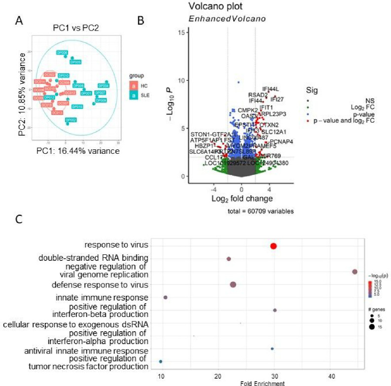 Figure 1
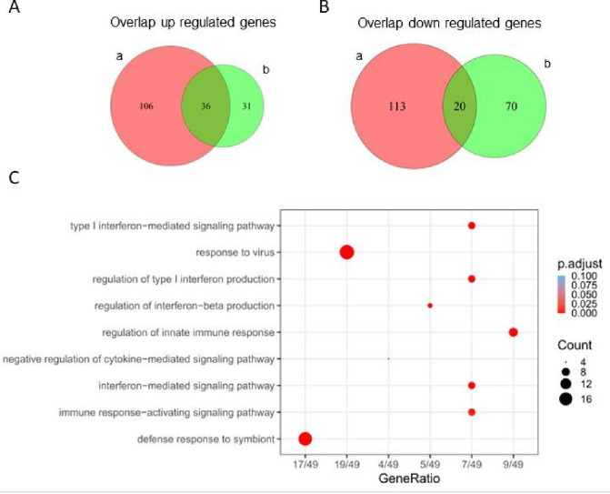 Figure 2
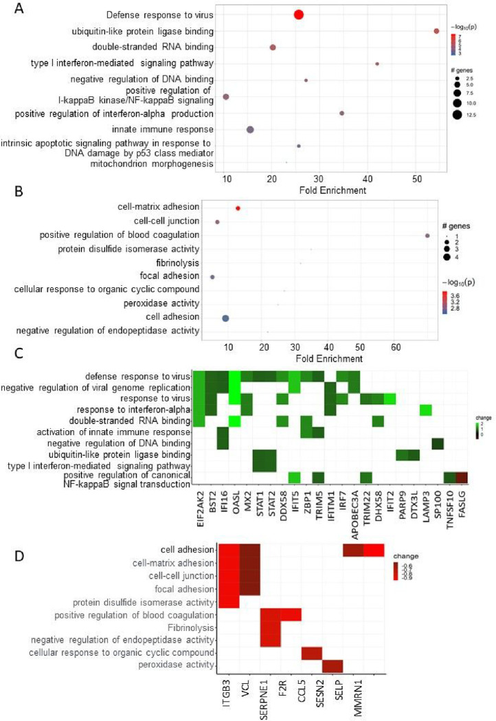 Figure 3
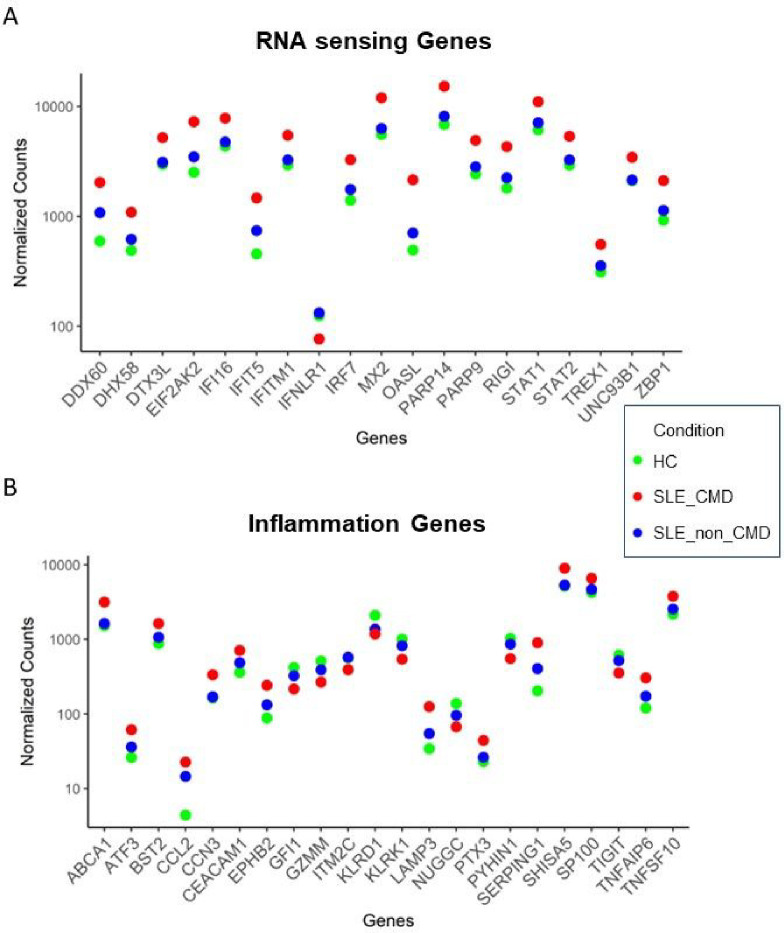 Figure 4
Peer Reviews
No public reviews on file for this paper yet. If you reviewed it on a platform where reviews are public (OpenReview, ICLR, NeurIPS, ICML), you can paste yours below so the community can read it here.
Videos
No videos yet. Explain this paper in a talk, walkthrough, or lecture? Add one.
Taxonomy
TopicsSystemic Lupus Erythematosus Research · Atherosclerosis and Cardiovascular Diseases · Viral Infections and Immunology Research
Introduction
Patients with SLE were over three times more likely to die of cardiovascular disease than patients in the general population[1]. Many women with SLE frequently report chest pain in the absence of obstructive coronary artery disease (CAD) due to coronary microvascular dysfunction (CMD), a form of ischemia with no obstructive CAD. CMD is a heart condition where blood flow response is impaired, resulting in reduced coronary flow reserve (CFR)[2]. This is associated with increased resistance in small blood vessels, spasms, limited myocardial perfusion reserve, and potential heart muscle ischemia, despite minimal blockage in the main heart arteries (less than 50% narrowing or a fractional flow reserve over 0.80)[3]. In terms of risk factors for CMD in the general population, notable associations have been found in women who have impaired coronary flow reserve with age, hypertension, smoking history, elevated heart rate, and low HDL[4]. Chronic inflammation also plays an important role in the pathophysiology of CMD[5]. For example, elevated levels of CRP, VCAM-1, PAI-1, vWF have been reported in CMD patients[6]. However, the molecular mechanisms underlying the development of CMD in SLE patients is unknown.
Cardiac magnetic resonance imaging (cMRI) is a non-invasive technology that allows for analysis of cardiac function, including CMD. cMRI measures determine the mass and volumes of the heart, as well as provide structural imaging of the myocardial tissue to detect fibrosis. Stress perfusion imaging facilitates the assessment of coronary blood flow, enabling the detection of ischemia and valve disorders. The myocardial perfusion index (MPRI) is a semi-quantitative method that demonstrates both sensitivity and specificity in diagnosing CMD, with an MPRI of less than 1.84 indicating CMD. Additionally, measures of left ventricular diastolic volumes and ejection fraction are employed to detect cardiac dysfunction. Previous studies by our group have demonstrated that approximately 40% of SLE patients have CMD on cMRI [7–9]. Interestingly in our most recent study we compared cardiac function by cMRI with clinical and inflammatory markers in a cohort of 13 SLE women[9]. We found that left ventricular function and cardiac strain were impaired in patients with SLE compared to reference controls and correlated with increased SLICC damage index and CRP levels. However, when we analyzed cardiac function and markers of inflammation in patients with and without CMD, we observed that patients without CMD contributed more to the observed differences between SLE and reference control groups, perhaps driven by increased left ventricular mass in patients without CMD versus patients with CMD. This prompted us to apply additional analyses to gain a better understanding of the molecular mechanisms underpinning CMD in SLE. RNA-sequencing (RNA-seq) offers valuable insights into the gene expression patterns within the transcriptome. In this study, our objective was to evaluate whether distinctive gene signatures can differentiate between SLE patients with CMD (SLE-CMD) and those without (SLE-non-CMD) by examining gene expression in healthy controls, SLE-CMD, and SLE-non-CMD whole blood. The findings presented in this article establish connections between peripheral biomarkers and the underlying pathobiology of SLE-CMD.
Results
Clinical information of the study subjects is described in our previous analysis[9]. All study subjects are female, with no significant differences in age or subject characteristics (such as BMI, fasting glucose or insulin levels, and inflammatory markers such as C3, C4, CRP and ESR) between groups. Whole blood transcriptomes from the cohort of 11 SLE patients (4 with CMD and 7 without CMD) and 10 age matched healthy controls (HC) were analyzed by RNA-sequencing (RNA-seq).
Principal Component Analysis (PCA) unveiled distinctive expression profiles between HC and SLE (Figure 1A). After data normalization using DEseq2, preprocessing and filtering with the criteria of padj <0.05, We identified 143 differentially expressed (DE) genes when comparing the SLE group and HC group. The DE gene analysis delineated a discernible molecular signature between SLE and HC, as displayed in Figure 1B. Upon further filtering with a |log2FC|>0.5 threshold, we identified 52 upregulated genes and 50 downregulated genes. Among the top 10 upregulated genes were IFI27, IFI44L, RSAD2, IFI44, SIGLEC1, IFIT1, SLC12A1, RPL23P3, CTXN2, ISG15, while the top 10 downregulated genes were RN7SKP227, RN7SL1, NTN4, RN7SL653P, SLC1A7, SGCD, S1PR5, KIR2DL3, LIM2, MMP23B. To gain insights into the functionality of those genes, we conducted a comprehensive Gene Ontology (GO) analysis. As expected, GO analysis results revealed SLE is significantly associated with gene enrichment of functions related to defense response to virus, type I interferon signaling pathway, and response to virus in upregulated DE genes (Figure 1C)[12–15].
Next, to understand if there were differences in gene signatures between patients with or without CMD, we conducted a DE analysis comparing SLE-CMD to SLE-non-CMD. Due to the considerable variability among the samples and coupled with the fact that the dataset size is relatively small, a direct comparison between SLE groups revealed only 14 DE genes at padj < 0.1 (table 1). To comprehensively understand if there were differences within whole blood transcriptomes between SLE-CMD, SLE-non-CMD, and HC groups, we employed the HC as reference and conducted a comparative analysis of commonly up regulated and down regulated genes between SLE-CMD and SLE-non-CMD. This generated datasets that comprised genes commonly upregulated or downregulated between SLE-CMD and SLE-non-CMD versus healthy control and genes uniquely up or down regulated in both patient subgroups (as shown by the Venn diagrams in figure 2A, B). Our investigation unveiled 36 genes that were consistently upregulated and 20 genes that were downregulated across both SLE-CMD and SLE-non-CMD groups when compared to the HC group (overlapping area of figure 2A and B and table 2). Furthermore, we identified 176 genes displaying unique expression patterns between SLE-CMD and HC (left hand area of Venn diagrams in figure 2A and B), along with 144 unique DE genes between SLE-non-CMD and HC (right hand area of Venn diagrams in figure 2A and B). Analyzing the DE genes in common between SLE-CMD and SLE-non-CMD, Gene Ontology (GO) and pathway enrichment analyses indicated that these genes are clearly associated with antiviral immune responses and were primarily IFN stimulated genes (Figure 2C).
We next conducted Ontology (GO) and pathway enrichment analyses for genes unique to SLE-CMD and SLE-non-CMD patients. Sorting by P value, the top GO terms of the biological process (BP), cellular component (CC) and molecular function (MF) categories are shown in Figure 3A (SLE-CMD) and 3B (SLE-non-CMD). As GO terms for RNA sensing, double stranded (ds) RNA and single stranded (ss) RNA binding, were enriched in SLE-CMD patient samples, we further analyzed the expression of the leading edge genes in the top GO terms (Figure 3C). In contrast only downregulated genes in SLE-non-CMD patients were associated with any GO categories. For example, genes associated with blood coagulation, cell-cell junction, and cellular defense response were decreased in SLE-non-CMD blood samples (Figure 3D).
Analyzing a subset of genes relevant to RNA sensing, we observed that a panel of genes associated with IFN signaling and response to RNA and DNA sensing such as RIGI, DDX60, DHX58 and ZBP1 were significantly increased in SLE-CMD compared to HC and SLE-non-CMD (Figure 4A, B). In contrast, down regulated genes included the inhibitory receptor TIGIT, and markers of natural killer cells (NK) or invariant NK T cells (iNKT) KLRG1 and KLRC, suggesting that cells and pathways that might restrain cardiotoxic responses are decreased in CMD patient blood samples. Another interesting finding was the observation that IFNLR1, a component of the receptor for IFN lambda, was decreased in SLE-CMD compared to SLE-non-CMD and HC. Enzymes involved in ADP-ribosylation, a post-translational modification that regulates protein function, are also differentially upregulated in SLE-CMD patients compared to non-CMD. PARP9 and PARP14 were both increased and are part of a sub family of ADP-ribosylation enzymes that recognize mono-ADP-ribosylation (MAR) on proteins as opposed to poly-ADP-ribosylation (PAR). However, in patients with non-CMD, pathways relative to coagulation, platelet activity and cell adhesion are decreased specifically relative to SLE-CMD patients. These results suggest that RNA sensing pathways are associated with development of vascular changes associated with CMD in SLE, whereas reduced platelet activity and coagulation is associated with the reduced left ventricular function we observed in the non-CMD cohort in our previous cardiac MRI study [9]. This is the first study to identify gene signatures that may associate with SLE.
Discussion
Chest pain is a common complaint among individuals with SLE even when there is no evidence of obstructive coronary artery disease (CAD)[8]. It is crucial to consider ischemic mechanisms like Coronary Microvascular Dysfunction (CMD) and coronary vasospasm during the diagnostic assessment. Given the heightened risk of cardiovascular disease (CVD) mortality and morbidity associated with SLE, stemming from inflammatory and metabolic pathophysiological processes, there is a pressing need for early identification and prevention of CVD risk factors. Hence, comprehending the molecular patterns within SLE-CMD may offer valuable insights into diagnostic methods and potential treatment approaches for clinical applications.
Endothelial dysfunction is a key driver of microvascular dysfunction and CMD. Endothelial cells (ECs) serve as a barrier between circulating blood components and the vascular wall and as such, play an important role in regulating immune responses to danger associated (such as oxLDL, heatshock proteins) and pathogen-associated molecular patterns (bacterial and viral proteins or nucleic acids) (DAMPs and PAMPs, respectively). In doing so they secrete cytokines and chemokines that regulate inflammation and immune responses, in addition to proteins that regulate vascular tone and function. Viral infection of ECs results in the activation of anti-viral signaling pathways that are activated to contain and eliminate the infection. One of the keyways viruses are detected by an infected cell is through recognition of viral RNA and DNA in the cytosol by RNA and DNA sensing proteins. RIG-I, MDA-5 and DHX58 (or LGP2) are three members of the RIG-I like receptors (RLRs) that are RNA helicases that recognize viral-derived dsRNA, become activated and drive the production of type I IFNs. A number of cardiotoxic agents such as 25-hydroxylcholesterol, angiotensin II and type I IFNs themselves have been shown to upregulate RIG-I expression. Together the increased expression of IFNs and IFN stimulated genes such as ISG15, CXCL10 and OAS2, are known to trigger EC dysfunction, specifically nitric oxide (NO) decreases, leukocyte adhesion, inflammation, coagulation, and endothelial cell injury. However, the direct role of RIG-I-mediated IFN induction in endothelial cells is relatively unexplored. One study showed that systemic administration of a RIG-I ligand led to impaired endothelium-dependent vasodilation in mouse aorta, although whether the effects of RIG-I activation were endothelial specific in this case was not shown. Our data clearly shows an association between upregulation of RIG-I, DHX58 and other components of RNA/DNA sensing such as ZBP2, in whole blood RNA samples from SLE patients with CMD compared to either healthy control subjects or SLE patients with chest pain but no evidence of CMD or obstructive disease. In addition, PARP9 and PARP14 have previously been shown to positively regulate signaling via RNA sensing pathways and IFNβ induction, strengthening the link with CMD pathobiology and RNA sensing [24-26]. This data would suggest that increased levels of genes involved in RNA sensing may contribute to enhanced expression of ISGs such as OAS2 and CXCL10 that can alter endothelial function and potentially contribute to CMD.
Another important finding in our study highlights the role of adaptive immune responses in SLE-CMD. Specifically, the observations that elevated levels of NKG2D associate with SLE-CMD, suggest a role for natural killer cells in immune events contributing to CMD. Relative to this study, studies have indicated that the interaction between immune cells and cardiomyocytes via NKG2D and its ligand NKG2DL can induce cardiomyocyte death, exacerbating cardiac remodeling following myocardial infarction (MI)[21]. The reduction of the T cell specific inhibitory receptor TIGIT in SLE-CMD compared to SLE-non-CMD whole blood samples, also supports a role for dysregulated adaptive immune responses. Exhausted T cells express TIGIT and are not only a feature of tumor immunity but also of SLE and lupus nephritis in the MRL/lpr model of SLE[22]. Our finding in SLE-CMD that TIGIT levels are decreased suggests that there is less of an exhausted phenotype in our cohort of SLE-CMD compared to SLE-non-CMD patients and that potentially T cell activity is contributing to microvascular defects.
In conclusion this study has unveiled a RNA sensing gene signature in whole blood samples of SLE patients with CMD compared to SLE patients without. Whilst our study is limited by the small samples size our signature is robust and agrees with recent literature examining the role of RIG-I and TREX1 in heart disease. While circulating microRNAs have been suggested as biomarkers for early-stage CAD, to date no biomarkers for CMD have been described. Both oxidative stress and epigenetic regulation of such pathways have previously been linked with CMD. Whether mitochondrial function and oxidative stress are contributing to mtRNA and DNA release in SLE with CMD patient immune cells is a potential mechanism driving these findings is an intriguing possibility [27, 28]. Interestingly, CMD is common in patients with prior COVID infection and CMD considered a strong driver of morbidity and mortality associated with COVID-19 [29, 30]. However, whether prior viral infection drove this signature in our CMD patient cohort is not known. Further analysis is required to understand the relationship between this gene signature, CMD and immune cell dysregulation.
Materials And Methods
Patients and Healthy Controls:
This study was approved by the institutional review board at Cedars-Sinai Medical Center and in line with the principles of the Declaration of Helsinki. All participants provided informed consent before joining. Participants were recruited from the lupus clinic. Inclusion criteria comprised female SLE subjects aged 37 to 57 years at baseline who experienced chest pain suspected to be angina. Exclusion criteria involved individuals with documented obstructive CAD and those with contraindications to coronary computed tomography angiography (CCTA) or cardiac magnetic resonance imaging (cMRI) as previously described [7]. Healthy controls were age matched and recruited from Cedars-Sinai Medical Center. blood samples were collected from all patients and controls in the fasting state using PAXgene blood RNA tubes.
PAXgene RNA isolation:
Blood samples were kept in PAXgene RNA tubes in −80 until ready to process. RNA was isolated using the PAXgene Blood RNA kit according to the manufacturers guidelines (PreAnalytiX). Eluted RNA was dissolved in RNase-free water. The quality and quantity of RNA were evaluated using the Agilent 2100 BioAnalyzer (Santa Clara, CA, USA).
RNA Sequencing:
Total RNA samples were analyzed for RNA integrity on the 2100 Bioanalyzer using the Agilent RNA 6000 Nano Kit (Agilent Technologies, Santa Clara, CA) and quantified using the Qubit RNA HS Assay Kit (ThermoFisher Scientific, Waltham, MA). Three hundred ng of total RNA Total was ribodepleted using the RiboCop Depletion Kit Human/Mouse/Rat v2 (Lexogen Inc., Greenland, NH). Stranded RNA-Seq library construction was performed using the xGen Broad-Range RNA Library Prep Kit (Integrated DNA Technologies, Coralville, IA). Library concentration was measured with a Qubit fluorometer (ThermoFisher Scientific), and library size was evaluated on a 4200 TapeStation (Agilent Technologies). Multiplexed libraries were sequenced on a NovaSeq 6000 (Illumina, San Diego, CA) using 75bp single-end sequencing. On average, approximately 50 million reads were generated from each sample.
Bioinformatics and data analysis:
Raw reads obtained from RNA-Seq were aligned to the transcriptome using STAR (version 2.5.0) /RSEM (version 1.2.25) with default parameters, using a custom human GRCh38 transcriptome reference downloaded from https://www.gencodegenes.org, containing all protein coding and long non-coding RNA genes based on human GENCODE version 33 annotation. Expression counts for each gene in all samples were normalized by a modified trimmed mean of the M-values normalization method and the unsupervised principal component analysis (PCA) was performed with DESeq2 Bioconductor package version 1.42.0 in R version 4.3. Each gene was fitted into a negative binomial generalized linear model, and the Wald test was applied to assess the differential expressions between two sample groups by DESeq2. Benjamini and Hochberg procedure was applied to adjust for multiple hypothesis testing, and differential expression gene candidates were selected with a false discovery rate less than 0.05. For functional enrichment analysis across sample groups, we conducted genes enrichment analysis using the R package “clusterProfiler v4.10.0”[10] and “pathfindR v2.3.1[11].
The reference list from the paper itself. Each links out to its DOI / PubMed record.
- 1Ajeganova S., Hafstrom I., and Frostegard J., Patients with SLE have higher risk of cardiovascular events and mortality in comparison with controls with the same levels of traditional risk factors and intima-media measures, which is related to accumulated disease damage and antiphospholipid syndrome: a case-control study over 10 years. Lupus Sci Med, 2021. 8(1).10.1136/lupus-2020-000454 PMC 787134533547230 · doi ↗ · pubmed ↗
- 2Del Buono M.G., , Coronary Microvascular Dysfunction Across the Spectrum of Cardiovascular Diseases: JACC State-of-the-Art Review. J Am Coll Cardiol, 2021. 78(13): p. 1352–1371.34556322 10.1016/j.jacc.2021.07.042PMC 8528638 · doi ↗ · pubmed ↗
- 3Taqueti V.R. and Di Carli M.F., Coronary Microvascular Disease Pathogenic Mechanisms and Therapeutic Options: JACC State-of-the-Art Review. J Am Coll Cardiol, 2018. 72(21): p. 2625–2641.30466521 10.1016/j.jacc.2018.09.042PMC 6296779 · doi ↗ · pubmed ↗
- 4Mygind N.D., , Coronary Microvascular Function and Cardiovascular Risk Factors in Women With Angina Pectoris and No Obstructive Coronary Artery Disease: The i POWER Study. J Am Heart Assoc, 2016. 5(3): p. e 003064.27068634 10.1161/JAHA.115.003064 PMC 4943278 · doi ↗ · pubmed ↗
- 5Godo S., , Role of Inflammation in Coronary Epicardial and Microvascular Dysfunction. Eur Cardiol, 2021. 16: p. e 13.33897839 10.15420/ecr.2020.47PMC 8054350 · doi ↗ · pubmed ↗
- 6Hung O.Y., , Novel biomarkers of coronary microvascular disease. Future Cardiol, 2016. 12(4): p. 497–509.27291585 10.2217/fca-2016-0012 PMC 5941701 · doi ↗ · pubmed ↗
- 7Hagiwara A.M., , Reduced left ventricular function on cardiac MRI of SLE patients correlates with measures of disease activity and inflammation. med Rxiv, 2023.10.26502/jrci.2809088 PMC 1094941338505536 · doi ↗ · pubmed ↗
- 8Manchanda A.S., , Coronary Microvascular Dysfunction in Patients With Systemic Lupus Erythematosus and Chest Pain. Front Cardiovasc Med, 2022. 9: p. 867155.35498009 10.3389/fcvm.2022.867155 PMC 9053571 · doi ↗ · pubmed ↗