From Data to Design: Constructing Scapula and Hip Bone Through Online Datasets, Open-Source Software and 3D Printers
Dharam S Rathia, Vijay K Konuri

TL;DR
This paper shows how to create detailed 3D-printed models of human bones using free software and CT scans, offering a cost-effective alternative to traditional models.
Contribution
The novel approach combines open-source tools and CT data to produce anatomically accurate 3D-printed bones for educational use.
Findings
3D-printed scapula and hip bone models retained most anatomical features with high accuracy.
Manual editing improved the clarity of morphological features in printed models.
The method proved effective for educational purposes despite minor limitations in fine details.
Abstract
Introduction Human skeletons and bones are essential for medical and allied sciences students. Nowadays, it is becoming challenging to procure bone legally, resulting in medical students' inadequacy of bone. Plaster-of-Paris or resin bone models sold on the market are less detailed than real bones. Aims and objectives This study aims to create a three-dimensional (3D)-printed human bone model with free, open-source software and fused deposition modelling (FDM) 3D printers, compare its accuracy with the original bones and validate it with a textbook description. Methods Scapula and hip bone models were produced using open-source software 3D Slicer from computed tomography (CT) data from the “New Mexico Decedent Image Database”. After automated segmentation, bones were edited manually with tools in a 3D Slicer (https://www.slicer.org/) and Meshmixer software (Autodesk Inc., San…
Genes, proteins, chemicals, diseases, species, mutations and cell lines named across the full text — each resolved to its canonical identifier and authoritative record.
Click any figure to enlarge with its caption.
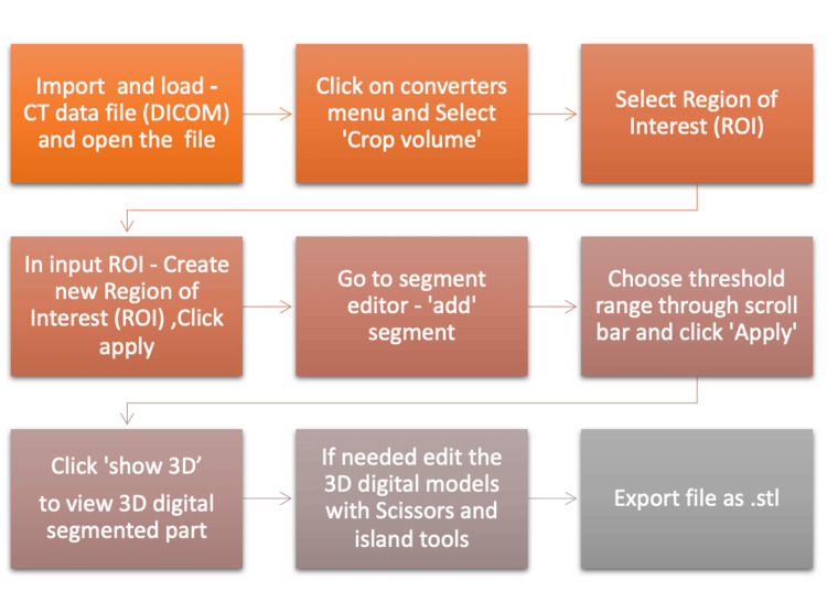 Figure 1
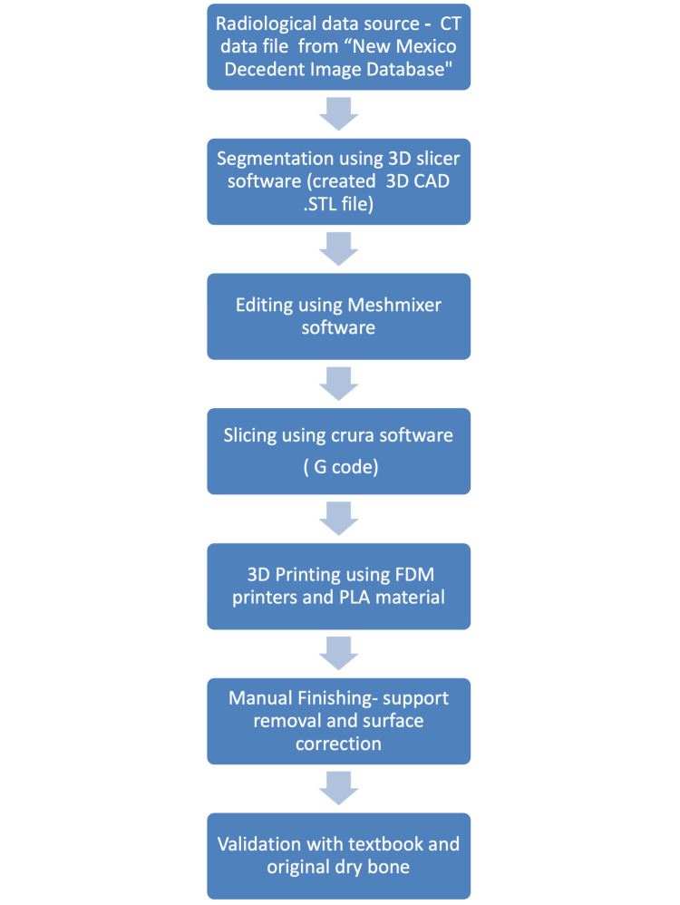 Figure 2
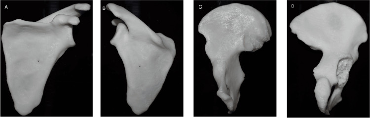 Figure 3
| S. No. | CT image series description | Bone | Sex | 3D Slicer threshold range, Max-Min |
| 1 | Thin bone HD-UXT | Scapula | Male | 128-2976 |
| 2 | Thin ST torso | Hip bone | Male | 232-2976 |
| Model | Original dimension of bone in milimeters (mm) | Dimension optimised for printing uniform scaling in milimeters (mm) | Print time in hours | Material used (in grams) | PLA filament length in meters (m) | ||||
| x | y | z | x | y | z | ||||
| Scapula | 119.603 | 96.733 | 172.463 | 120 | 80.14 | 57.905 | 6.15 | 53 | 17.69 |
| Hip bone | 126.365 | 136.293 | 246.635 | 130 | 78 | 60.8 | 7.9 | 49 | 16 |
| S. No. | List of anatomical features of scapula | Anatomical accuracy of digital model (.stl) | Anatomical accuracy of 3D printed physical model after post-processing |
| 1 | Ventral surface | Accurate few spikes like projection | Accurate |
| 2 | Dorsal surface | Accurate small, hole | Accurate |
| 3 | Spine | Accurate | Accurate |
| 4 | Coracoid process | Accurate | Accurate |
| 5 | Acromion | Accurate | Accurate |
| 6 | Glenoid fossa | Surface with attached humerus | Accurate |
| 7 | Axillary margin | Accurate | Accurate |
| 8 | Medial angle | Accurate few spikes | Accurate |
| 9 | Vertebral margin | Accurate | Accurate |
| S. No. | Anatomical features of hip bone or innominate | Anatomical accuracy of digital model (.stl) | Anatomical accuracy of 3D printed physical model after post-processing | |
| 1 | Ilium | Iliac crest | Accurate | Accurate |
| Greater sciatic notch | Accurate | Accurate | ||
| Lesser sciatic notch | Accurate | Accurate | ||
| Iliac tuberosity | Accurate | Accurate | ||
| Preauricular sulcus | Accurate | Accurate | ||
| Iliac fossa | Accurate | Accurate | ||
| Auricular surface | Obscured with attachment of sacrum | Accurate | ||
| Anterior superior and inferior spines | Accurate | Accurate | ||
| 2 | Ischium | Ischial tuberosity | The surface feature not appreciable | Accurate |
| 3 | Pubis | Pubic symphyses | Accurate | Accurate |
| 4 | Structures formed by the intersection of the three bones of the innominate: | Obturator foramen | Accurate | Accurate |
| Acetabulum Lunate fossa | Obscured by the head of the humerus need digital and manual correction after printing | Fragment of bone remains in the fossa not clearly appreciable | ||
Peer Reviews
No public reviews on file for this paper yet. If you reviewed it on a platform where reviews are public (OpenReview, ICLR, NeurIPS, ICML), you can paste yours below so the community can read it here.
Videos
No videos yet. Explain this paper in a talk, walkthrough, or lecture? Add one.
Taxonomy
TopicsAnatomy and Medical Technology · Shoulder Injury and Treatment · Hip disorders and treatments
Introduction
Human skeletons and bones are essential for medical and allied sciences students [1-3]. In the early 2000s, the buying and selling of human skeletal material were prohibited in many countries [4]. Since 1985, the Indian government has imposed export prohibitions following a thorough analysis of the illegal bone trade [5]. The legal market fails to meet the demand, resulting in the use of illicit sources [6]. Even medical colleges in India have bone sets, and they are inadequate for a large number of students [2]. To address the ethical concerns and inadequacy of human bone, alternatives such as three-dimensional (3D) images, plastic models and wooden skull models have been explored as substitutes for human bones in teaching anatomy [6-8]. Plaster-of-Paris, or resin bone models that are sold in the market, are not as detailed as those of real bones [9]. Innovations like the Skeleton Virtual reality application have also been introduced to enhance students' learning and engagement in studying anatomy, but lack haptic feelings [10]. One helpful tool for modernising anatomy education is the creation of 3D-printed models that are anatomically accurate [11]. 3D printing or additive manufacturing (AM) is the layer-by-layer creation of a physical object from a 3D computer model [12]. Detailed methodologies have been developed to create optimized models of bone for 3D printing [13,14]. Here, we provide a similar method of making a 3D-printed anatomical model of the human scapula and innominate or hip bone using the open-source software 3D Slicer (https://www.slicer.org/), Meshmixer (Autodesk Inc., San Rafael, California, United States), and fused deposition modelling (FDM) 3D printers to observe the accuracy of gross anatomical features. This study aims to create a 3D-printed human bone model with free, open-source software and FDM 3D printers, compare its accuracy with the original bones and validate it with a textbook.
Materials and methods
Radiological data source
Computed tomography (CT), Digital Imaging and Communications in Medicine (DICOM) data file, the international standard for medical images and related information of the individual were downloaded from the "New Mexico Decedent Image Database" [15].
Creating 3D model using 3D Slicer
The segmentation of CT scan data of a patient from the database, image series for the upper limb region and torso, was done using open-source 3D Slicer software (Version 6.6.2) [16]. At first, we segmented the scapula using the thin bone "H.D.-UXT CT" image series. Before segmentation, the new region of interest (ROI) was selected with a converter menu and crop volume. Then, segmentation was done by the segment editor by selecting the threshold range 128-2976 (Figure 1; Table 1).
Process of image segmentation using 3D Slicer software from CT dataImage created by Rathia DS and Konuri VK.
Segmented regions were visualised by clicking the "Show 3D" icon. The individual left side scapula was isolated with editing tools like an island and a scissor in the segment editor. After segmentation, part of the humerus remains attached to the bone, and the surface of the bone shows a few unwanted spikes and holes. These parts were removed using an editing tool in 3D Slicer and Meshmixer software. Finally, the stereolithography or standard triangulation language (.stl) file of the bone model was exported and saved. The innominate or hip bone was segmented using the "Thin S.T. torso" CT image series. Like scapula segmentation, the new ROI was created with a converter menu and crop volume. Then, with the segment editor, manually segmented the left hip bone by selecting threshold range 232-2976 (Table 1, Figure 1). The "Show 3D" icon was used to visualise a segmented region in a 3D view. An individual hip bone was isolated from the ROI using an island and scissor tool. The .stl file of bone was exported and saved. Then, the file is inspected and corrected using Meshmixer software (Version 3.5.474). Further editing of both bone models was done with Meshmixer software to smoothen the articulating surface and remove the head of the humerus, femur and sacrum attached.
Creating physical 3D models using FDM 3D printers
The .stl file was converted into a geometric code (G codes) file using a 3D Slicer Crura software (version 16.04.6.). Models were optimised and reduced in size for printer-specific requirements (Table 2).
Then, with the help of a 3D computer model, it was converted into a physical object using the FDM or fused filament fabrication (FFF) 3D printer (Pratham 3.0; Make3d.in, Surat, India). Using commercially available common 3D printing materials, polylactic (PLA) 3D models were created at 80% infill density. Post-processing, like surface smoothening and support removal, was done using a hand sander and drill. The models were compared with original dry bones and text descriptions from the anatomy book [17]. The following flowchart shows the whole process of our study flow chart of study (Figure 2).
Flow chart showing the step-by-step process of creating 3D modelsImage created by Rathia DS and Konuri VK.FDM: fused deposition modelling; PLA: polylactic filament
Results
With the help of 3D Slicer software, we segmented the scapula and hip bone. The model of the scapula obtained lacks anatomical accuracy over the surface. A few morphological features can not be segmented like the original dry bone, and a few finer details over the surface of the supraspinous and interspinous fossa were uneven. The anatomical features of the final 3D scapula digital and physical models printed with a 3D printer were similar to the original dry bone and feature mentioned in the textbook (Figure 3; Table 3) [17].
3D printed bone of scapula: (A) anterior view, (B) posterior view; Hip bone: (C) medial view, (D) lateral viewImage created by Rathia DS and Konuri VK.
The model of hip bone obtained too had few faint anatomical features over the gluteal surface and ischium when compared with dry bones. A few morphological features differed from the original dry bone, and a few finer details were unclear. After segmentation, the articulating surface was not visible in the acetabular cavity. Part of the head of the femur and part of the sacrum remain attached to the bone, and the bone shows a few unwanted spikes and holes. These parts were removed using an editing tool in 3D Slicer and Meshmixer software. Finally, the anatomical features of the hip bone 3D digital model obtained after editing and physical models printed with a 3D printer were similar to the original dry bone and feature mentioned in the textbook (Table 4) [17].
Discussion
Research summary
Bone models of the scapula and hip bone were produced using open-source software 3D Slicer from CT data obtained from the "New Mexico Decedent Image Database" [15]. 3D Slicer is a free, open-source software for visualising, processing, segmenting, registering, and analysing medical, biomedical, and other 3D images and meshes and planning and navigating image-guided procedures [16]. Although the software used in the study is not meant for clinical use, it helps segment anatomical regions for research and education [16]. Segmentation is the division of magnetic resonance (MR) or CT images of an organ into distinct anatomical structures or segments of different tissue types done manually and automated [18]. After automated segmentation, bones were edited manually with tools in a 3D Slicer and Meshmixer and 3D prints were printed using PLA filament. PLA is cost-effective, is generally easier to work with, and produces more aesthetic-looking parts [19].
Interpretation of findings
We aimed to test the capability of free and open-source software to create bone models. After a few minor edits, the outcome showed that the artificial bone scapula and innominate created were anatomically as accurate as real dry bones described in the textbook. The surface and features were edited using tools in 3D Slicer and Meshmixer. The final physical model obtained after manual post-processing had most of the features and was accurate. The 3D digital skeleton of the hip bone retained all anatomical features except articulating surfaces, such as the acetabulum and auricular surface ilium, which were obscured by the head of the femur and sacrum. We found this method a potential alternative solution for creating accurate human bones. A study used and contrasted three distinct methods - 3D scanning, photogrammetry, and micro-CT - to create a digital 3D zygomatic bone and micro-CT was found to be the most effective method for achieving morphological accuracy [11]. A similar study utilizing the same free software, the 3D Slicer and the New Mexico Decedent Image Database successfully generated precise models of 2D human bones of the foot [13]. 3D-printed anatomical models were created to study frogs for anatomy education after 3D scanning and printing skeletal tissues [20]. As per the ethical concern and inadequacy that appeared in the various research articles, accurate bone in an adequate number can be produced using 3D printing bone models; it is an ethically potential solution for medical students [1,2,4,6]. Many studies have already mentioned the utility of 3D printed models for anatomy education for improving student engagement and skeletal tissue for medical and biology education [3,20,21].
Limitations and implications
Although we successfully created anatomically accurate artificial bones, it is limited by a few unreproduced surface features. These can be resolved by editing with individual experts in computer-assisted design (CAD) designing, and more accurate models can be produced as we desire pathological models. With 3D segmentation and printing, apart from creating more units of bone models for students' demonstration, we can also produce rare anatomical models with variations and pathologies, accidental fractures, or the investigation of skeletal remains.
Conclusions
In summary, this study investigates the method of creating 3D-printed human bone scapula and hip bone models. We successfully created 3D digital models with a 3D Slicer with maximum possible accuracy, and a few incorrect anatomical features in digital models were corrected using Meshmixer software. It was possible to produce anatomically accurate models that bore the greatest possible resemblance to the original bones and the textbook descriptions after 3D printing and post-processing. These models could be alternatives to real dry bones in medical colleges where sufficient bones are unavailable. This method could utilised by clinicians to create patient-specific models for medical education and research.
The reference list from the paper itself. Each links out to its DOI / PubMed record.
- 1Med students go hunting for skeletons 4 2024 2016 https://bangaloremirror.indiatimes.com/bangalore/others/med-students-go-hunting-for-skeletons/articleshowprint/54554094.cms
- 2The bioethics of skeletal anatomy collections from India Nat Commun Agarwal SC 16921520243840220010.1038/s 41467-024-45738-6PMC 10894195 · doi ↗ · pubmed ↗
- 3Emphasise details of 3D-printed bones with contrast paints Med Educ Poór VS Tóth D Simon G 1915720233646046110.1111/medu.14985 PMC 10351435 · doi ↗ · pubmed ↗
- 4Human Tissue Act 2004 Human Tissue Acthttps://www.legislation.gov.uk/ukpga/2004/30/contents
- 5Government bans export of human skeletons 3 2024 2014 https://www.indiatoday.in/magazine/economy/story/19851130-government-bans-export-of-human-skeletons-802181-2014-01-20
- 6Anatomists' uses of human skeletons: ethical issues associated with the India bone trade and anonymized archival collections Anat Sci Educ Jones DG 6106171620233703930910.1002/ase.2280 · doi ↗ · pubmed ↗
- 7The wooden skull: an innovation through the use of local materials and technology to promote the teaching and learning of human anatomybio Rxiv Mugagga K Mwarisi MG Dare SS 202010.1155/2020/8036737 PMC 747435332908914 · doi ↗ · pubmed ↗
- 8Three-dimensional plastic modeling on bone frames for cost-effective neuroanatomy teaching Cureus Ramirez MJ Nurmukhametov R Musa G 014202210.7759/cureus.27472 PMC 942110236060355 · doi ↗ · pubmed ↗