Complete genome sequence of Microbacterium paraoxydans phage Damascus
Julisa M. Bearhart, Jenna L. Bethke, Cassie S. Christian, Faith N. Cour, Karleigh R. Creasey, Emily J. Crowe, Julia G. Dahl, Lindsey A. Hanson, Abby L. Jaecks, Vincent A. Lamantia, Mercedes Madison, Autumn L. Roskowiak, Justin D. Scheberl, Bekkah M. VanEperen, Morgan E. Wurst

TL;DR
This paper presents the full genome sequence of the Damascus phage, which infects Microbacterium paraoxydans and was found in Wisconsin soil.
Contribution
The study provides a new phage genome sequence and its classification within cluster EL.
Findings
Damascus has a 56,477 bp genome with 3′ single-stranded overhangs and 56.5% G+C content.
It belongs to cluster EL and shares 42.6%–91.7% gene content with other phages in this cluster.
Abstract
Phage Damascus was isolated from soil in northwestern Wisconsin using Microbacterium paraoxydans as the host. The Damascus genome is 56,477 bp with 3′ single-stranded overhangs and 56.5% G+C content. Damascus was assigned to cluster EL and shares 42.6%–91.7% gene content with the three other phages in this cluster.
Genes, proteins, chemicals, diseases, species, mutations and cell lines named across the full text — each resolved to its canonical identifier and authoritative record.
Click any figure to enlarge with its caption.
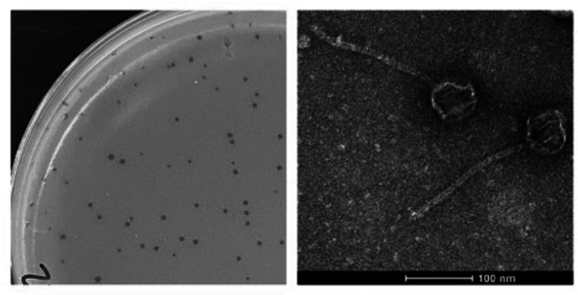 Fig 1
Fig 1Peer Reviews
No public reviews on file for this paper yet. If you reviewed it on a platform where reviews are public (OpenReview, ICLR, NeurIPS, ICML), you can paste yours below so the community can read it here.
Videos
No videos yet. Explain this paper in a talk, walkthrough, or lecture? Add one.
Taxonomy
TopicsBacteriophages and microbial interactions · Genomics and Phylogenetic Studies · Microbial infections and disease research
ANNOUNCEMENT
Comparative analysis of the genomes of phages infecting a single host genus, such as Microbacterium, has provided important insights into bacteriophage evolution and diversity (1). Here, we report the genome sequence of phage Damascus, isolated on Microbacterium paraoxydans NRRL B-14843. Damascus was isolated from soil collected 12 September 2022 in Amery, WI (45.311936 N, 92.366226 W) using standard procedures (2). Briefly, soil was suspended in peptone-yeast extract-calcium (PYCa) liquid medium and incubated with shaking at 250 rpm for 2 h at 30°C. The wash was collected by centrifugation and filtration (0.22-μm pore size), and the filtrate was inoculated with an overnight culture of Microbacterium paraoxydans. Following incubation with shaking for 48 h at 30°C, the culture was filtered, and the filtrate was plated in PYCa top agar with Microbacterium paraoxydans using three rounds of plaque purification. Plates were incubated for 24 h at 30°C. Plaques were clear and 1–2 mm in diameter. Negative stain transmission electron microscopy revealed that Damascus has siphoviral morphology, with an isometric capsid and a long, flexible tail (Fig. 1).
Characteristics of phage Damascus. (Left) Representative plaques formed by phage Damascus on Microbacterium paraoxydans; (right) high-titer lysates were placed on Formvar-coated grids, negatively stained with 1% uranyl acetate (2) and imaged using a FEI Tecnai Spirit BioTwin transmission electron microscope at 120 kV. Damascus has an average head diameter of 62 nm and tail length of 196 nm (n = 6).
Double-stranded DNA was isolated from phage lysate using the Promega Wizard DNA cleanup system, and a sequencing library was prepared using the NEB Ultra II kit. Sequencing was performed using an Illumina MiSeq system (v.3 reagents), yielding 471,080 150-bp single-end reads (1,197-fold genome coverage). Raw reads were assembled using Newbler (v.2.9), and completeness was verified using Consed (v.29.0) (3). The Damascus genome is 56,477 bp with nine-base 3′ single-stranded overhangs (CGCGTCACT) and 56.6% G+C content.
The genome was annotated using DNA Master (http://cobamide2.bio.pitt.edu), PECAAN (https://discover.kbrinsgd.org), GLIMMER (v.3.02) (4), GeneMark (v.2.5) (5), Starterator (v.1.1) (http://phages.wustl.edu/starterator/), and Phamerator (6). Gene functions were predicted using BLASTp using the National Center for Biotechnology Information (NCBI) nonredundant database (7) and Actinobacteriophage Database (8), HHpred (9) using the PDB mmCIF70, Pfam-A, and NCBI Conserved Domain databases, and DeepTMHMM (10). Genes encoding tRNAs were identified using ARAGORN (v.1.2.38) (11) and tRNAscan-SE (v.3.0) (12). Default settings were used for all programs. Annotation identified 82 protein-coding genes and no tRNA genes. We were able to assign predicted functions for 23 gene products (28%). Two gene products (gp40 and gp66) have no homologs in the database (8). Damascus is predicted to have a lytic life cycle based on the absence of genes associated with lysogeny.
Damascus was assigned to cluster EL, based on gene content similarity (GCS) of 35% or higher with phages in the Actinobacteriophage Database (8), as described (13). Within cluster EL, Damascus shares 91.7% GCS with DizzyRudy (MK814756), which was isolated on the same strain of M. paraoxydans, but only 72.4% and 42.6% with Camille (MH153800) and Count (MH153801), respectively, isolated on Microbacterium aerolatum B-24229. Damascus shares notable genome features with other cluster EL phages and with phages in several clusters (AM, AU, AW, BI CC, and DJ) isolated on different host genera (1). These features include a fused major capsid subunit and capsid maturation protease (gp14), two major tail proteins (gp17 and gp19), and relatively low percent G+C content compared to the host bacteria (56.5% for Damascus vs 67% for Microbacterium hosts) (1).
The reference list from the paper itself. Each links out to its DOI / PubMed record.
- 1Jacobs-Sera D, Abad LA, Alvey RM, Anders KR, Aull HG, Bhalla SS, Blumer LS, Bollivar DW, Bonilla JA, Butela KA, et al.. 2020. Genomic diversity of bacteriophages infecting Microbacterium spp. P Lo S ONE 15:e 0234636. doi:10.1371/journal.pone.023463632555720 PMC 7302621 · doi ↗ · pubmed ↗
- 2Poxleitner M, Pope W, Jacobs-Sera D, Sivanathan V, Hatfull GF. 2018. HHMI SEA-PHAGES phage discovery guide. Available from: https://seaphagesphagediscoveryguide.helpdocsonline.com/home
- 3Russell DA. 2018. Sequencing, assembling, and finishing complete bacteriophage genomes. Methods Mol Biol 1681:109–125. doi:10.1007/978-1-4939-7343-9_929134591 · doi ↗ · pubmed ↗
- 4Delcher AL, Bratke KA, Powers EC, Salzberg SL. 2007. Identifying bacterial genes and endosymbiont DNA with glimmer. Bioinformatics 23:673–679. doi:10.1093/bioinformatics/btm 00917237039 PMC 2387122 · doi ↗ · pubmed ↗
- 5Besemer J, Borodovsky M. 2005. Gene Mark: web software for gene finding in prokaryotes, eukaryotes and viruses. Nucleic Acids Res 33:W 451–W 454. doi:10.1093/nar/gki 48715980510 PMC 1160247 · doi ↗ · pubmed ↗
- 6Cresawn SG, Bogel M, Day N, Jacobs-Sera D, Hendrix RW, Hatfull GF. 2011. Phamerator: a bioinformatic tool for comparative bacteriophage genomics. BMC Bioinformatics 12:395. doi:10.1186/1471-2105-12-39521991981 PMC 3233612 · doi ↗ · pubmed ↗
- 7Altschul SF, Gish W, Miller W, Myers EW, Lipman DJ. 1990. Basic local alignment search tool. J Mol Biol 215:403–410. doi:10.1016/S 0022-2836(05)80360-22231712 · doi ↗ · pubmed ↗
- 8Russell DA, Hatfull GF. 2017. Phages DB: the actinobacteriophage database. Bioinformatics 33:784–786. doi:10.1093/bioinformatics/btw 71128365761 PMC 5860397 · doi ↗ · pubmed ↗