Case Report: an unusual orbital tumor
Anis Mahmoud, Hager Touil, Fadima Hann, Riadh Messaoud, Amira Trigui, Mehdi Hasnaoui, Anis Mahmoud

TL;DR
A rare case of orbital lipoma in a 63-year-old patient is reported, highlighting its clinical presentation, diagnostic challenges, and successful treatment.
Contribution
This case report adds to the limited literature on orbital lipomas and emphasizes the importance of histology for accurate diagnosis.
Findings
The patient presented with diplopia and limited elevation of the right eye due to a fusiform fatty lesion in the inferior rectus muscle.
Complete tumor excision via a transconjunctival approach led to full recovery with no recurrence after 3 years of follow-up.
Histopathological confirmation was essential for diagnosis, as clinical evaluation alone was insufficient.
Abstract
Introduction Orbital lipoma is an extremely rare tumor, representing less than 1% of all orbital tumors. We review the literature and describe the presentation, the differential diagnosis and the management of this tumor. Case report We report the case of a 63-year-old patient who was referred for a diplopia with recent hemi-cranial headache. Physical examination showed no exophthalmos nor decrease in visual acuity. The patient complained of diplopia on elevation and oculomotricity examination showed limited elevation of the right eye. The Hess Lancaster test was in favor of a limited course of the right inferior rectus muscle. Magnetic resonance imaging revealed a fusiform tissue process in the right inferior rectus muscle with a fatty signal. A complete excision of the tumor was performed by a trasncunjonctival approach. Cytopathological examination was consistent with a…
Genes, proteins, chemicals, diseases, species, mutations and cell lines named across the full text — each resolved to its canonical identifier and authoritative record.
Click any figure to enlarge with its caption.
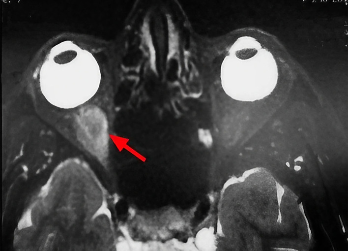 Figure 1
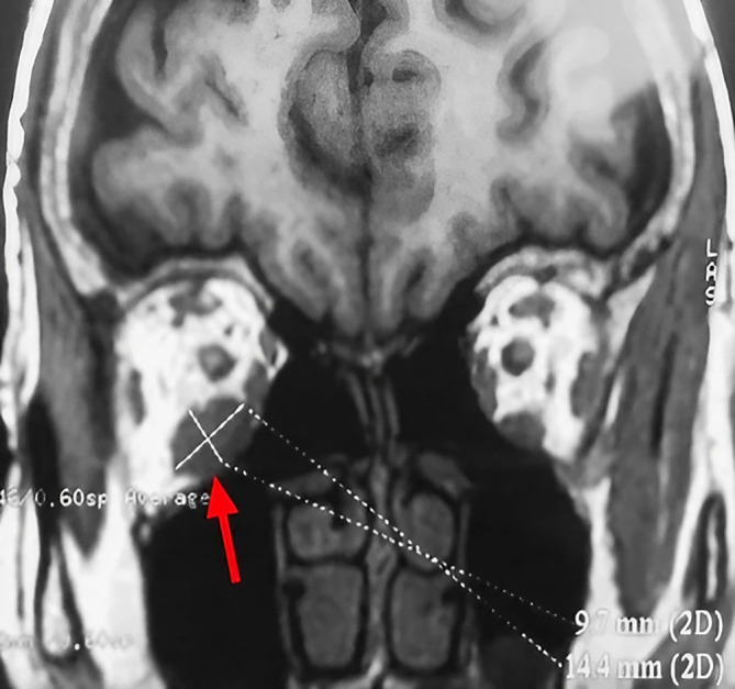 Figure 2
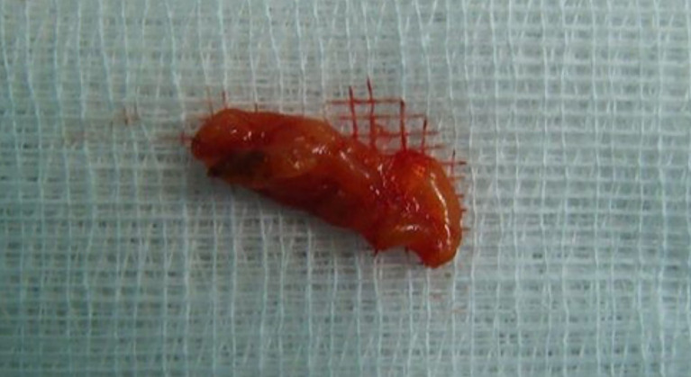 Figure 3
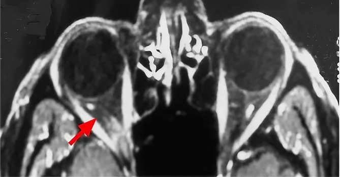 Figure 4
Peer Reviews
No public reviews on file for this paper yet. If you reviewed it on a platform where reviews are public (OpenReview, ICLR, NeurIPS, ICML), you can paste yours below so the community can read it here.
Videos
No videos yet. Explain this paper in a talk, walkthrough, or lecture? Add one.
Taxonomy
TopicsSoft tissue tumor case studies · Sarcoma Diagnosis and Treatment · Cancer and Skin Lesions
Introduction
Lipomas are benign tumors, rare in the orbit, representing less than 1% of all orbital tumors. They pose a differential diagnosis with a variety of other expansive orbital masses. ^ 1 ^ We report a new case, review the literature and discuss the clinicopathological and radiological features, the differential diagnosis and the management of this entity.
Case report
A 63-year-old unemployed Tunisian woman, with no previous personal or family pathological history, presented with a diplopia evolving for two weeks. Physical examination showed no exophthalmia and no decrease in visual acuity. Furthermore, it revealed diplopia on elevation. Oculomotricity examination showed limited elevation of the right eye, which was confirmed by the Hess Lancaster test that revealed a limited course of the right inferior rectus muscle. Magnetic resonance imaging (MRI) showed a fusiform and hyper-vascularized tissue process located in the right inferior rectus with fatty signal. The tumor was hyperintense on spin-echo T2-weighted images ( Figure 1) and hypointense on spin-echo T1-weighted images ( Figure 2).
Sagittal section of orbital MRI showing a mass with high signal on a T2 weighted image (red arrow).
Coronal section of orbital MRI showing a mass with low signal on a T1 weighted image (red arrow).
These findings suggested various diagnosis; lipoma, inflammatory process, lymphoma and malignant tumor.
We performed a right inferior transconjunctival orbitotomy and excisional biopsy under general anesthesia. Peroperatively, we discovered an encapsulated mass of fatty tissue, thus complete excision was made. No adherences or involvement of adjacent structures occurred. The specimen was well circumscribed and slightly firmer than normal adipose tissue, with a yellow surface ( Figure 3).
Macroscopic appearance of the specimen.
Histologic examination was consistent with a pleomorphic lipoma. The postoperative period was uneventful. Immediately after the operation, the patient reports the resolution of his diplopia. Postoperative MRI images demonstrated the complete resolution of the tumor ( Figure 4). With 3 years of follow up, there is no sign of recurrence or ocular motility impairment.
Postoperative axial MRI image demonstrating complete tumor resolution (red arrow).
Discussion
Orbital lipoma is the most common mesenchymal soft tissue tumor. However, it is rarely found in the orbit despite the presence of abundant adipose tissue in the intraorbital space. ^ 2 ^ ^,^ ^ 3 ^ A review of the largest series of orbital tumors revealed a very low incidence of lipomas. ^ 4 ^ Shields et al. reported only two cases of lipomas in a review of 1264 cases of orbital tumors, indicating the rarity of this entity. ^ 5 ^ On physical examination, the diagnosis is often difficult to suggest. These tumors are often asymptomatic.
However, they can cause severe morbidity by causing progressive and painless exophthalmos, which is occasionally coupled with diplopia or ocular motility defects ^ 6 ^ such as was observed in our patient.
Orbital lipoma exceptionally leads to a compressive neuropathy responsible for a significant decrease in visual acuity, an alteration of the afferent photomotor reflex and the visual field constriction. ^ 1 ^ Imaging based on computed tomography (CT) scanning and MRI is essentially useful in ascertaining determining the exact seat, size and relationship to the orbit content. The fatty signal is characteristic on CT sequences. Furthermore, as was found in our patient, the tumor is hypointense on spin-echo T1-weighted images and hyperintense on spin-echo T2-weighted images. ^ 1 ^
Histology is essential for definitive diagnosis of pleomorphic lipoma. An important histologic criterion is the presence of a mixture of fat cells, pleomorphic cells and in particular floret-like multinucleated giant cells embedded in a myxoid stroma. ^ 7 ^ That concorded with the histological result of our case. Differential diagnosis of this tumor became more important because the number of reports about some other tumors of similar morphology, are increasing. Pleomorphic lipoma may be confused with lipomatous hemangiopericytoma, myofibroblastoma or even malignant tumors such as rhabdomyosarcoma, myxoid malignant fibrous histiocytoma and liposarcoma. ^ 8 ^ ^,^ ^ 9 ^ Surgical excision of an orbital lipoma is not only recommended for symptomatic cases such as our patient's clinical presentation but also to exclude malignancy. ^ 10 ^ In addition, as was noted in our patient, the long-term outcome after surgery is considered excellent. ^ 11 ^
This case highlights the importance of orbital imaging in the context of diplopia without obvious cause to rule out an intraorbital lipoma. Nevertheless, this association remains rare and requires further documentation of cases.
Conclusion
Lipomas are benign soft tissue tumors, rarely located in the orbit. The clinical presentation is variable depends on the size and the intraorbital site. The histology makes the definitive diagnosis and may precisely identify the variant.
Consent
Written informed consent for publication of their clinical details and/or clinical images was obtained from the patient.
The reference list from the paper itself. Each links out to its DOI / PubMed record.
- 1Chi MJ Roh JH Lee JH : A case of orbital lipoma with exophtlmos and visual disturbance. Jpn. J. Ophtalmol. 2009;53(4):442–444. 10.1007/s 10384-009-0679-2 19763768 · doi ↗ · pubmed ↗
- 2Civit T Klein O Freppel S : Tumeurs orbitaires d’origine mésenchymateuse. Neurochirurgie. 2010;56:158–164. 10.1016/j.neuchi.2010.02.007 20227093 · doi ↗ · pubmed ↗
- 3Tripathy D Mittal R : Spindle cell lipoma of the orbit. Ophthal. Plast. Reconstr. Surg. 2015;31:e 53–e 55. 10.1097/IOP.0000000000000068 24819207 · doi ↗ · pubmed ↗
- 4Kim MH Sa HS Woo K : Fibrolipoma of the orbit. Ophthal. Plast. Reconstr. Surg. 2011;27:e 16–e 18. 10.1097/IOP.0b 013e 3181 dee 5fb 20700070 · doi ↗ · pubmed ↗
- 5Shields JA Shields CL Scartozzi R : Survey of 1264 patients with orbital tumors and simulating lesions: The 2002 montgomery lecture, part 1. Ophthalmology. 2004;111:997–1008. 10.1016/j.ophtha.2003.01.002 15121380 · doi ↗ · pubmed ↗
- 6Toledano FN Stoica BT Genol SI : Diplopia from pleomorphic lipoma of the orbit with lateral rectus muscle involvement. Ophthalmic Plast. Reconstr. Surg. 2013;29(2):e 53–e 55. 10.1097/IOP.0b 013e 31826 a 5112 23328779 · doi ↗ · pubmed ↗
- 7Bartley GB Yeatts RP Garrity JA : Spindle cell lipoma of the orbit. Am. J. Ophtalmol. 1985;100:605–609. 10.1016/0002-9394(85)90691-9 4050936 · doi ↗ · pubmed ↗
- 8Ulivieri S Oliveri G Motolese PA : Spindle cell lipoma of the orbit: acase report of an unusual orbital pathology. Neurol. Neurochir. Pol. 2010;44:419–423. 10.1016/S 0028-3843(14)60303-0 20827617 · doi ↗ · pubmed ↗