Unresolved haemosporidia of the Australian skink, Egernia stokesii
Kristína Zechmeisterová, Michael George Gardner, Pavel Široký

TL;DR
This study investigates the haemosporidian parasites in the Australian skink Egernia stokesii, revealing conflicting results between molecular and morphological analyses.
Contribution
The study highlights the challenges in identifying haemosporidian species due to conflicting molecular and morphological data.
Findings
Molecular data clustered isolates with Haemocystidium, but morphology resembled Plasmodium.
Limited sampling and indistinguishable nuclei/merozoites hindered species identification.
Mixed infections may be present but cannot be confirmed with current data.
Abstract
The Australian skink Egernia stokesii had been recognised as a host of two species of Plasmodium, Plasmodium mackerrasae and P. circularis; nevertheless, molecular data are available for only a single haemosporidian species of this host. Its sequences are labelled as “Plasmodium sp.” or “Plasmodium mackerrasae”, but morphological characteristics of this isolate are unavailable. Phylogenetic analyses of these sequences placed them into the clade of the genus Haemocystidium. In this study, blood samples of six E. stokesii were analysed by both, molecular and microscopic methods to clarify the haemosporidia of this lizard. Application of these approaches offered discordant results. Whereas sequence analysis clustered our isolates with lizard species of Haemocystidium, morphology of blood stages is more akin to Plasmodium than Haemocystidium. However, limited sampling, indistinguishable…
Genes, proteins, chemicals, diseases, species, mutations and cell lines named across the full text — each resolved to its canonical identifier and authoritative record.
Click any figure to enlarge with its caption.
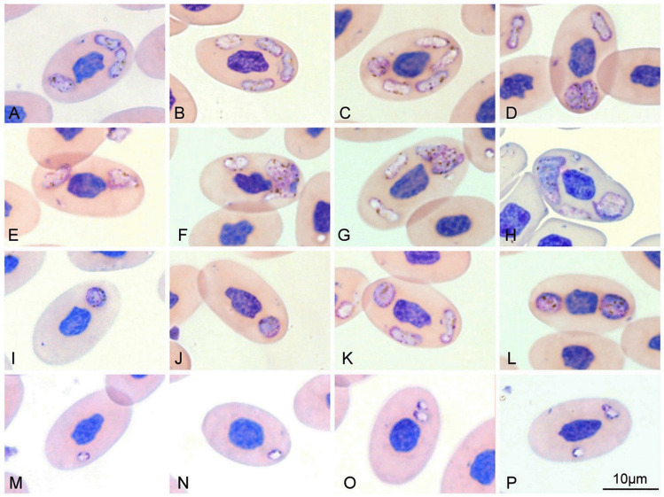 Figure 1
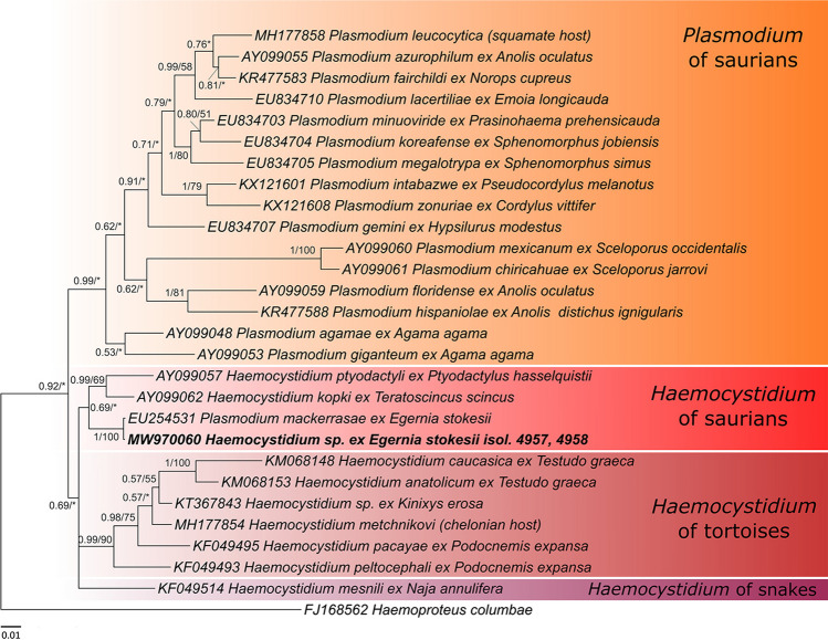 Figure 2
- —University of Veterinary Sciences Brno
- —University of Veterinary Sciences Brno
Peer Reviews
No public reviews on file for this paper yet. If you reviewed it on a platform where reviews are public (OpenReview, ICLR, NeurIPS, ICML), you can paste yours below so the community can read it here.
Videos
No videos yet. Explain this paper in a talk, walkthrough, or lecture? Add one.
Taxonomy
TopicsBird parasitology and diseases · Vector-borne infectious diseases · Amphibian and Reptile Biology
Introduction
Genera Plasmodium and Haemocystidium represent well-known blood protistan parasites of reptiles distributed worldwide (Telford 2009; Pineda-Catalan et al. 2013; Maia et al. 2016; O’Donoghue 2017; Galen et al. 2018). Plasmodium is the most common haemosporidian genus containing more than one hundred species described from lizards. On the other hand, only fifteen species of Haemocystidium were reported from lizards (Telford 1982, 2009; Paperna and Landau 1991; Telford et al. 2012).
Sequences labelled “Plasmodium sp.” and “Plasmodium mackerrasae” available in GenBank are known from the Australian skink Egernia stokesii (Gray 1845). Whilst Martinsen et al. (2008) labelled four sequences obtained in their study as Plasmodium sp., one of these sequences (EU254531) appeared as Plasmodium mackerrasae later in article by Galen et al. (2018) and another one (EU254574) already with changed generic allocation as Haemocystidium sp. in article by Boysen et al. (2022). However, these sequences did not cluster with lizards’ Plasmodium species, but instead within the Haemocystidium clade (Martinsen et al. 2008; Galen et al. 2018). Since these sequences are not accompanied with morphological data, their taxonomic delimitations have not yet been characterised (Martinsen et al. 2008; Galen et al. 2018). Egernia stokesii is a known host for both Plasmodium and Haemocystidium species as well as the haemogregarine Hemolivia mariae Smallridge et Paperna 1997 (Kvičerová et al. 2014).
Two Plasmodium species have been described from E. stokesii: Plasmodium mackerrasae Telford 1979, and Plasmodium circularis Telford et Stein 2000 (Telford 1979; Telford and Stein 2000). According to Telford (2009), another seven haemosporidian species are known from Australian lizards. Plasmodium australis Garnham 1966 infecting eastern bearded dragon Pogona barbata (Cuvier 1829) (formerly Amphibolurus barbatus), Plasmodium billbraya (Paperna et Landau 1990) from the gecko Christinus marmoratus (Gray 1845), Plasmodium egerniae Mackerras 1961 was found in the skink Bellatorias major (Gray 1845) (formerly Egernia major). Notably, P. egerniae was never detected from E. stokesii during 3-year study carried out by J. Stein (Telford 2009). Four species of Haemocystidium were identified from Australian geckos: Haemocystidium gehyrae (Paperna et Landau 1991) described from Gehyra australis Gray 1845, Haemocystidium mackerrasae (Paperna et Landau 1991) (original spelling mackerrasi) from Heteronotia binoei (Gray 1845), Haemocystidium oedurae (Paperna et Landau 1991) from Oedura castelnaui (Thominot 1889), and Haemocystidium underwoodsauri (Paperna et Landau 1991) from Underwoodisaurus milii (Bory de Saint-Vincent 1825) (Paperna and Landau 1991; Telford 2009). A recent diversity-focussed study by Boysen et al. (2022) dealt with haemosporidians and haemogregarines of lizards across northern tropical Australia. Using sequence analysis, authors found new evolutionary lineages of Haemocystidium in geckos of the genera Oedura and Gehyra. Nevertheless, Egernia skinks were not included in their collection and their study did not provide any nomenclatoric outcomes.
The aim of our study was to characterise haemosporidia detected in E. stokesii by both molecular and microscopic methods. Obtained data were compared to the available information to attempt a species identification for the haemosporidia found in E. stokesii.
Materials and methods
Six individuals of E. stokesii were captured during September and October 2012 at the southern Flinders Ranges, South Australia. Blood smears and Whatman® FTA® cards-preserved blood samples were processed as is described in Kvičerová et al. (2014). Microscopic examination of the Giemsa-stained blood smears was carried out under an Olympus BX-53 microscope; photomicrographs of parasite stages was taken with an Olympus DP 73 digital camera and QuickPhoto Micro 3.0 software at × 1000 magnification with immersion oil. Maximum length and width of parasite stages were measured and LW (length × width) and L/W (length/width) values were calculated. All measurements were in micrometres (μm) unless stated otherwise and are presented as the mean ± standard deviation and range (in parentheses). Parasitaemia was estimated as the proportion of infected cells per 10^4^ examined erythrocytes, expressed as a percentage (%).
Genomic DNA was extracted from blood samples using the DNeasy Blood & Tissue Kit (Qiagen, Hilden, Germany) according to the manufacturer´s instructions as was described by Kvičerová et al. (2014). Haemosporidian parasites were detected by amplification of cyt b gene using three nested primer sets according to previous studies. Primers DW2/DW4 and DW1/DW6 amplify a 1200 bp fragment (Perkins and Schall 2002) and HaemnF1/HaemnR3 and HaemF/HaemR2 amplify a shorter—480 bp fragment of various haemosporidians, including genera Haemocystidium and Plasmodium (Bensch et al. 2000; Hellgren et al. 2004; Javanbakht et al. 2015; Oliveira et al. 2018). To distinguish between Plasmodium and Haemoproteus/Haemocystidium we used a third nested PCR with combination of primers AE298/AE299 (outer) and AE983/AE985 (inner, 558 bp) and AE980/AE982 (inner, 346 bp), respectively, according to Pacheco et al. (2018) amplifying only Plasmodium. Co-infection by haemogregarines was detected by EF/ER primers according to Kvičerová et al. (2008). All PCR reactions were carried out in a 25 μl volume with Combi PPP Master Mix (Top-Bio s.r.o, Prague, Czech Republic). Both negative (PCR water) and positive (DNA of Haemocystidium anatolicum and Plasmodium relictum) controls were included in PCR reactions. Positive PCR products were purified using a Gel/PCR DNA Fragments Extraction Kit (Geneaid Biotech, New Taipei City, Taiwan) and bidirectionally Sanger sequenced by service laboratory (Macrogen Inc., Amsterdam, the Netherlands). Sequence and phylogenetic analyses were performed in Geneious 11.0.5 software (Kearse et al. 2012; http://www.geneious.com). Our sequences were compared to all available sequences by BLAST algorithm (Altschul et al. 1990). Tree-alignment created by the MUSCLE algorithm (Edgar 2004) consisted of lizards’ Plasmodium and reptiles’ Haemocystidium species with Haemoproteus columbae (FJ168562) as an outgroup. A phylogenetic tree was calculated by two methods—Bayesian inference (BI) with plugin Mr. Bayes 3.2.6. (Huelsenbeck and Ronquist 2001) and Maximum likelihood (ML) with plugin Phyml 3.3.2 (Guindon et al. 2010), both under GTR evolutionary model. The resulting trees were summarised in TreeGraph 2.15 (Stöver and Müller 2010) and graphically edited in Inkscape 0.92.4 (http://www.inkscape.org/).
Results
Microscopy
Two out of six blood smears were positive for haemosporidians with parasitaemia levels of 3.84% and 4.72%. Since Hemolivia infection was observed in all six blood smears, both haemosporidia positive blood smears were coinfected with Hemolivia mariae. All observed haemosporidian parasites were intraerythrocytic and multiple infections within one host cell were common. Four different morphologies of haemosporidian parasites were observed.
- The most common observed stage had an elongate shape and oval ends (Fig. 1A–D, F, G and K) with cytoplasm stained whitish purple and being darker at margins. Dark brown pigmented granules of haemozoin were present in their cytoplasm. Nuclei were not clearly visible. These stages were positioned randomly around the host cell nucleus.
- The biggest stages had an irregular, rectangular or lobate shape (Fig. 1E–H), with whitish purple cytoplasm speckled with many little dark brown pigmented granules of haemozoin. Dark purple or dark blue granules, resembling nucleus or nuclei of merozoites, occupied a large part of the cytoplasm (Fig. 1F, G, H). These stages were located mostly in polar position regarding to the host cell nucleus.
- Round to oval stages (Fig. 1I–L) possessed a darker purple cytoplasm, sometimes with dark blue granules resembling chromatin. Dark brown pigmented granules of haemozoin were present. These stages were located predominantly also in polar position regarding to the host cell nucleus.
- The smallest stages (Fig. 1M–P) had an oval shape, with whitish cytoplasm, and their dark blue nuclei were located at one pole. Few haemozoin granules were sometimes present. Fig. 1. Four different morphologies of haemosporidia observed in the blood smears from Egernia stokesii. Elongate stages A–C and D (upper); irregular stages E–H, suspected meront H (left); round stages I–L; and the smallest stages M–P. Various stages were observed within one red blood cell in D, F, G and K
Measurements of all stages are summarised in Table 1.Table 1. Morphological comparison of intraerythrocytic stages of Haemocystidium sp. from this study with Plasmodium mackerrasae**Haemocystidium sp. (this study)Plasmodium mackerrasaeTelford and Stein 2000Trophozoites: 1.87 ± 0.44 (1–3) × 1.52 ± 0.49 (1–2), N = 30Trophozoites: 1.5 × 0.8 to 2–3 × 1.0–1.5Stages of irregular shape with suspected multiple nuclei (meronts?)7.96 ± 0.82 (7–9) × 4.31 ± 0.47 (4–5), N = 3Meronts: 3 × 2 to 4 × 2.5 (binucleate); 4–5 × 2.5–4 (tetranucleate); 5.1 ± 1.1 × 3.7 ± 1.1 (3–7 × 2.5–6.0, N = 25) with 4–14 merozoites (mature);Elongate stages: 5.26 ± 0.64 (4–7) × 2 ± 0 (2–2), N = 30Immature gametocytes (elongate shape): 5–7 × 1.5–2.5Round stages: 4.08 ± 0.67 (3–6) × 3.46 ± 0.5 (3–4), N = 30Mature gametocytes (round shape): 5.8 ± 0.9 × 4.6 ± 0.7 (3.5–9.0 × 3.0–6.0, N = 75)Stages of irregular shape: 6.46 ± 1.23 (5–9) × 4.25 ± 0.64 (3–6), N = 30Chronic phase gametocytes (irregular shape): 6.1 ± 0.9 × 4.6 ± 0.5 (5.0–9.0 × 3.5–6.0, N = 25)
Phylogenetic analyses of sequence data
Corresponding to positive blood smears, sequences (cyt b) were obtained from both haemosporidia-positive blood samples. Sequence chromatograms were clear, no signal of mixed infection was found. All obtained sequences were identical; thus, only the two longest sequences (1012 and 1059 bp) were used in the phylogenetic tree. The longer sequence was submitted into the GenBank database under accession number MW970060. Our sequences were identical with Plasmodium sp. EU254531 (607 bp) from the same host E. stokesii (57% query coverage). In the phylogenetic tree (Fig. 2), our sequences and Plasmodium sp. EU254531 clustered within the Haemocystidium clade from saurian hosts with Haemocystidium ptyodactyli (Paperna et Landau 1991) from lizard Ptyodactylus hasselquistii (Donndorff 1798) from Israel (AY099057) and Haemocystidium kopki De Mello 1916 from Teratoscincus scincus (Schlegel 1858) from Pakistan (AY099062) as closest relatives. Haemocystidium species form their own monophylum, which appears to be divided into three clades regard to vertebrate host: (1) Haemocystidium spp. from lizards, (2) turtles and (3) snakes. Branching is not well supported, nevertheless, this branching is consistent with previous studies (Pineda-Catalan et al. 2013; Galen et al. 2018).Fig. 2. Phylogenetic tree based on Bayesian inference (BI) of cyt b of haemosporidians from Egernia stokesii. Topology from Mr. Bayes analysis is shown with nodal supports as BI posterior probabilities/ML (maximum likelihood) bootstrap values. Bootstrap values lower than 50 are marked with asterisk. Sequences obtained in this study are marked in bold
Hemolivia mariae infection was molecularly confirmed in all six blood samples.
Discussion
Sequence analyses placed the haemosporidian isolate from E. stokesii within a cluster of saurian Haemocystidium species and therefore, we propose it should be classified in the genus Haemocystidium. Furthermore, the identical sequence to ours, EU254531, originally classified by previous authors as Plasmodium sp. or “P. mackerrasae “ seems to be also Haemocystidium instead (Martinsen et al. 2008; Galen et al. 2018). Morphological data are not available for the sequence EU254531, so a morphological comparison with our samples is not possible. The other two Haemocystidium isolates of lizards with DNA sequences available in GenBank—Haemocystidium ptyodactyli and H. kopki—are morphologically different from the Haemosporidia that we observed. Haemocystidium ptyodactyli has a big halteridial gametocytes and H. kopki has huge round gametocytes (Telford 1982, 2009; Paperna and Landau 1991). In contrast to our findings, the remaining morphologically described lizards’ Haemocystidium species have significantly larger mature gametocytes, often occupying most of the erythrocyte’s cytoplasm, whereas suspected mature stages in our samples are relatively small. Furthermore, we did not observe a halteridial morphology, which is typical for the majority of described Haemocystidium species. Two Haemocystidium species described from Australian geckos, H. mackerrasae and H. underwoodsauri, have a smaller round microgametocyte, but only the early stages have a size similar to our isolates. Later, or mature gametocytes of H. mackerrasae and H. underwoodsauri have a larger size than our Haemocystidium species. Furthermore, H. mackerrasae possess a pair of chromatin bodies, which we did not observe, and H. underwoodsauri has a halteridial shape. Other round shaped Haemocystidium species—H. gehyrae, H. grahami Shortt 1922, H. kopki and H. tarentolae (Parrot 1927) are significantly larger and/or have a vacuolated cytoplasm (Telford 2009). Suspected trophozoites in our samples are morphologically similar to trophozoites of many Haemocystidium and Plasmodium species, and thus, they cannot be used in differential diagnosis*.*
The blood stages we observed in the blood of E. stokesii were morphologically most similar to P. mackerrasae (Telford and Stein 2000; Telford 2009). The elongated stages resemble immature gametocytes and the round stages mature gametocytes. The irregular stages were similar to the meronts, but also to the gametocytes in the chronic phase of the infection. Both mature meronts and gametocytes of P. mackerrasae in the chronic phase have irregular shapes but are similar in size, but can be distinguished by the presence of merozoites in the former. The presence of round stages, which are typical for active infections of P. mackerrasae, suggests that the irregular stages are likely meronts with merozoites. Unfortunately, merozoites or multiple nuclei, typical for the genus Plasmodium were not clearly distinguishable in the stages of irregular shape that we observed (suspected meront with merozoites in Fig. 1F, G, H). Measurements of the various stages of our Haemocystidium sp. and P. mackerrasae are comparable (Table 1). The presence of haemozoin pigment and mostly polar location of gametocytes and meronts also resemble P. mackerrasae. Plasmodium circularis, which is also known to infect E. stokesii possesses halteridial and dumbbell-shaped forms and is morphologically different from our findings (Telford and Stein 2000; Telford 2009).
Despite the morphological similarity of our haemosporidia with P. mackerrasae and possible presence of dividing stages, based on the molecular analysis it is classified as Haemocystidium. This genus, unlike Plasmodium, does not show dividing stages (meronts) in the peripheral blood. Nevertheless, multinucleate stages with 2–3 nuclei have been described in four Haemocystidium species of lizards; Haemocystidium papernai Telford 1996, Haemocystidium quettaensis Telford 1996, Haemocystidium lygodactyli Telford 2005 and Haemocystidium apigmentada Telford, Peirce, et Samour 2012 (Telford 2009; Telford et al. 2012). This trait is considered to be an aberration in early nuclear development of Haemocystidium gametocytes. In comparison, the meronts of P. mackerrasae have 4–14 merozoites (nuclei), and thus, it is unlikely to be an aberration in that case (Telford 1979; Telford and Stein 2000).
Interestingly, meronts of H. mariae with their oval to rounded shape, considerable size and vacuolisation (Kvičerová et al. 2014) resemble gametocytes of rounded Haemocystidium species. Meronts of H. mariae were observed in all the blood smears examined in this study, even in haemosporidia PCR-negative samples, but contained no haemozoin. We are therefore confident that we correctly distinguished between meronts of H. mariae and the observed haemosporidia stages.
Currently it is uncertain if the haemosporidia we have found is a Haemocystidium or a Plasmodium, or to which species belongs to. The same problem faced Galen et al. (2018), who suspected possibility of a mixed infection of Plasmodium and Haemocystidium, with PCR detecting only one parasite. However, we carried out numerous PCRs with subsequent sequence analyses and no signs of coinfection was detected. In morphology-based species descriptions we cannot exclude the possibility that mixed infection may have affected the original description. However, we have no indication of a similar problem in the case of P. mackerrasae (Telford 1979, 2009; Telford and Stein 2000). Despite the fact that some blood stages we observed more resemble the Haemocystidium sp*.* and some more resemble the Plasmodium sp., we conclude we were working with Haemocystidium morphologically resembling Plasmodium. It is also possible that we are working with a yet undescribed species. However, we do not think that we have currently enough unambiguous data to describe it as new. We suggest thorough sampling effort in populations of the previously mentioned host species to characterise all relevant Plasmodium and Haemocystidium species mentioned in this work by DNA barcoding, or whole genome sequencing, interconnected with species morphological characteristics. Applying such integrative approach, it will be possible to resolve the taxonomic conundrum of haemosporidia from E. stokesii.
The reference list from the paper itself. Each links out to its DOI / PubMed record.
- 1Altschul SF Gish W Miller W Myers EW Lipman DJ Basic local alignment search tool J Mol Biol 199021540341010.1016/S 0022-2836(05)80360-22231712 · doi ↗ · pubmed ↗
- 2Bensch S Stjernman M Hasselquist DÚstmanÚHansson B Westerdahl H Pinheiro RT Host specificity in avian blood parasites: a study of Plasmodium and Haemoproteus mitochondrial DNA amplified from birds Proc R Soc B 20002671583158910.1098/rspb.2000.118111007335 PMC 1690711 · doi ↗ · pubmed ↗
- 3Boysen KE Perkins SL Hunjan S Oliver P Gardner MG Balasubramaniam S Melville J Diversity and phylogenetic relationships of haemosporidian and hemogregarine parasites in Australian lizards Mol Phylogenet Evol 202216710735810.1016/j.ympev.2021.10735834774764 · doi ↗ · pubmed ↗
- 4de Oliveira JP AndréMR Alves Júnior JRF Lustosa APG Werther K Molecular detection of hemogregarines and haemosporidians in Brazilian free-living testudines Int J Parasitol Parasites Wildl 20187758410.1016/j.ijppaw.2018.01.00830050752 PMC 6058349 · doi ↗ · pubmed ↗
- 5Galen SC Borner J Martinsen ES Schaer J Austin CC West J Perkins SL The polyphyly of Plasmodium: comprehensive phylogenetic analyses of the malaria parasites (order Haemosporida) reveal widespread taxonomic conflict R Soc Open Sci 2018517178010.1098/rsos.17178029892372 PMC 5990803 · doi ↗ · pubmed ↗
- 6Guindon S Dufayard J-F Lefort V Anisimova M Hordijk W Gascuel O New algorithms and methods to estimate maximum-likelihood phylogenies: assessing the performance of Phy ML 3.0Syst Biol 20105930732110.1093/sysbio/syq 01020525638 · doi ↗ · pubmed ↗
- 7Hellgren O Waldenström J Bensch SA new PCR assay for simultaneous studies of Leucocytozoon, Plasmodium, and Haemoproteus from avian blood J Parasitol 20049079780210.1645/GE-184R 115357072 · doi ↗ · pubmed ↗
- 8Huelsenbeck JP Ronquist FMRBAYES: Bayesian inference of phylogenetic trees Bioinformatics 20011775475510.1093/bioinformatics/17.8.75411524383 · doi ↗ · pubmed ↗