A double-incision endoscopic myotomy of the cricopharyngeal muscle for the management of a large hemi-circumferential Zenker’s diverticulum
Sunil Gupta, Tony He, Jeffrey D. Mosko, Natalia Soledad Causada Calo, Christopher Teshima, Gary R. May

Abstract
Genes, proteins, chemicals, diseases, species, mutations and cell lines named across the full text — each resolved to its canonical identifier and authoritative record.
Click any figure to enlarge with its caption.
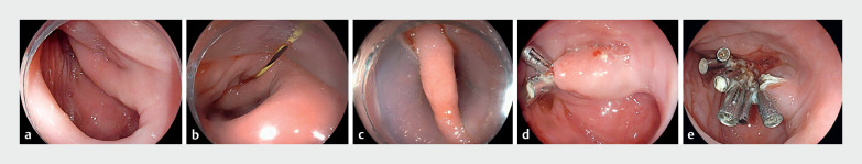 Fig. 1
Peer Reviews
No public reviews on file for this paper yet. If you reviewed it on a platform where reviews are public (OpenReview, ICLR, NeurIPS, ICML), you can paste yours below so the community can read it here.
Videos
No videos yet. Explain this paper in a talk, walkthrough, or lecture? Add one.
Taxonomy
TopicsDysphagia Assessment and Management · Esophageal and GI Pathology · Tracheal and airway disorders
An 81-year-old man with a decade-long history of dysphagia to solids presented to our hospital in extremis. He had lost 20 pounds in weight over 7 months, and more recently had been unable to tolerate clear fluids. A gastroscopy, barium swallow, and computed tomography scan revealed a large hemi-circumferential Zenker’s diverticulum, located 18 cm from the incisors and centered at 9 o’clock ( Fig. 1 a ), with minimal passage of contrast into the esophageal lumen observed.
Endoscopic images showing: a a large hemi-circumferential Zenker’s diverticulum located 18 cm from the incisors and centered at 9 o’clock; b a 0.035-inch guidewire placed to guide a Zenker’s overtube toward the diverticulum; c isolation of the muscular septum of the Zenker’s diverticulum; d the remnant posterior component of the Zenker’s diverticulum following completion of the first myotomy; e complete flattening of the Zenker’s diverticulum following completion of a second myotomy and clip closure.
Owing to a long muscular septum, we proceeded to perform a double-incision endoscopic dissection of the cricopharyngeal muscle ( Video 1 ). Zenker’s diverticulum peroral endoscopic myotomy (zPOEM) was not considered because of the potential need for two tunnels. After the diverticulum had been thoroughly cleaned, a 0.035-inch guidewire was placed into the stomach ( Fig. 1 b ). This was used to guide placement of a Zenker’s overtube (ZDO-22-30; Cook Medical, Bloomington, Indiana, USA) into the diverticulum to isolate the muscular septum ( Fig. 1 c ). A complete myotomy of the cricopharyngeal muscle was performed with a precut needle-knife (Zimmon knife; Cook Medical). After clip closure, re-evaluation revealed that the posterior component of the diverticulum remained prominent ( Fig. 1 d ) and there was still resistance to passage of the endoscope into the esophagus. The Zenker’s overtube was reinserted, isolating the remnant muscular septum. A second complete myotomy of the cricopharyngeal muscle was performed, followed again by clip closure. Subsequently to this, there was flattening of entire diverticulum and easy passage of the gastroscope into the esophagus ( Fig. 1 e ). Over the next few days, the patient’s dysphagia to liquids and solids completely resolved.
A double-incision Zenker’s myotomy is performed for the management of a large hemi-circumferential Zenker’s diverticulum.Video 1
Flexible endoscopic dissection of the cricopharyngeal muscle is well established as a safe and effective treatment option for a symptomatic Zenker’s diverticulum 1 . Our novel double-incision myotomy enabled complete flattening of a large hemi-circumferential diverticulum. In our experience, the use of a Zenker’s overtube aided exposure of the remnant septal bridge and facilitated the second myotomy. Further studies pertaining to the clinical outcomes of this approach are required.
Endoscopy_UCTN_Code_TTT_1AO_2AF
The reference list from the paper itself. Each links out to its DOI / PubMed record.