Fas2EB112: a tale of two chromosomes
Tara M Finegan, Christian Cammarota, Oscar Mendoza Andrade, Audrey M Garoutte, Dan T Bergstralh

TL;DR
This paper solves a genetic mystery about why two Fas2 mutations in fruit flies cause different effects by identifying a modifier mutation in the nrg gene.
Contribution
The study identifies Nrg14 as the modifier mutation responsible for enhancing the severity of the Fas2EB112 mutation.
Findings
The Fas2EB112 mutation's severe phenotype is enhanced by the Nrg14 mutation.
The Fas2G0336 mutation shows a mild phenotype due to suppression by a modifier.
Nrg14 is a classic null allele of the nrg gene, which encodes a cell-cell adhesion molecule.
Abstract
The cell–cell adhesion molecule Fasciclin II (Fas2) has long been studied for its evolutionarily conserved role in axon guidance. It is also expressed in the follicular epithelium, where together with a similar protein, Neuroglian (Nrg), it helps to drive the reintegration of cells born out of the tissue plane. Remarkably, one Fas2 protein null allele, Fas2G0336, demonstrates a mild reintegration phenotype, whereas work with the classic null allele Fas2EB112 showed more severe epithelial disorganization. These observations raise the question of which allele (if either) causes a bona fide loss of Fas2 protein function. The problem is not only relevant to reintegration but fundamentally important to understanding what this protein does and how it works: Fas2EB112 has been used in at least 37 research articles, and Fas2G0336 in at least three. An obvious solution is that one of the two…
Genes, proteins, chemicals, diseases, species, mutations and cell lines named across the full text — each resolved to its canonical identifier and authoritative record.
Click any figure to enlarge with its caption.
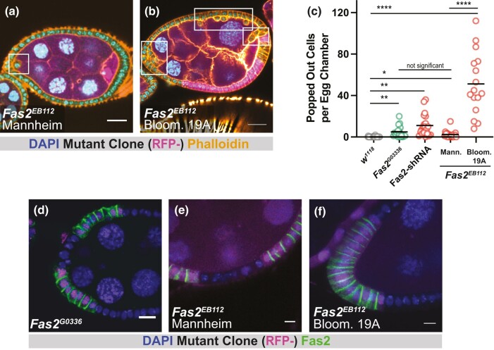 Fig. 1
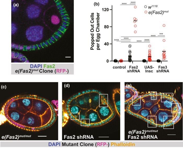 Fig. 2
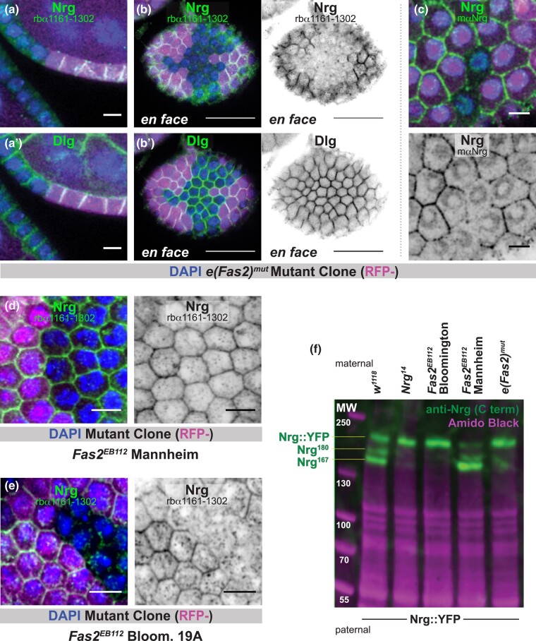 Fig. 3
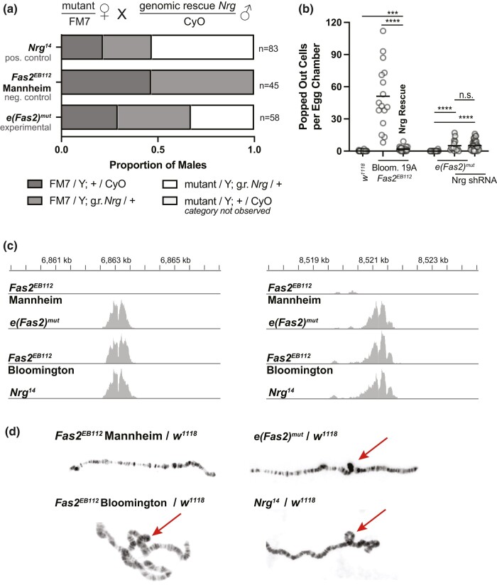 Fig. 4
- —National Institutes of Health10.13039/100000002
Peer Reviews
No public reviews on file for this paper yet. If you reviewed it on a platform where reviews are public (OpenReview, ICLR, NeurIPS, ICML), you can paste yours below so the community can read it here.
Videos
No videos yet. Explain this paper in a talk, walkthrough, or lecture? Add one.
Taxonomy
TopicsAxon Guidance and Neuronal Signaling · Nerve injury and regeneration · Microtubule and mitosis dynamics
Introduction
The Drosophila Immunoglobulin-superfamily cell adhesion molecules Fasciclin II (Fas2, orthologous to vertebrate NCAM) and Neuroglian (Nrg, orthologous to vertebrate L1-CAM), were identified in the developing nervous system, where they localize along fasciculating axons (reviewed in Harden et al. 2016). Functional studies have made extensive use of two experimentally generated null alleles. Fas2^EB112^, the first Fas2 allele, was made using imprecise excision of a P-element, resulting in a 1.7 kb deletion on the X chromosome that is thought to include the Fas2 transcriptional start site (Grenningloh et al. 1991). Nrg^14^ (previously called l(1)RA35), which was generated using X-ray mutagenesis, is an inversion that disrupts the Nrg gene sequence (Lefevre 1981; Hall and Bieber 1997). Early studies demonstrated that both proteins help to drive axon guidance and also that this function is evolutionarily conserved (reviewed in Araújo and Tear 2003). While their roles in the nervous system have received the most attention, Fas2 and Nrg have also been studied in a variety of developing epithelial tissues, including imaginal discs (Mao and Freeman 2009), Malphigian (renal) tubules (Halberg et al. 2016), the intestine (Resnik-Docampo et al. 2017), the follicular epithelium (Szafranski and Goode 2007; Gomez et al. 2012; Fic et al. 2021), and trachea (Neuert et al. 2020) (reviewed in Finegan and Bergstralh 2020).
One function shared by Fas2 and Nrg is epithelial cell reintegration. Many epithelial cells undergo a change in position during mitosis. Interkinetic nuclear migration, which is the apical-directed movement of the cell nucleus prior to division, is primarily studied in pseudostratified tissues (reviewed in Spear and Erickson 2012), but apical-directed mitotic cell movement is also evident in simple cuboidal and columnar epithelia, including the mammalian small intestine and ureteric bud (Sauer 1937; Packard et al. 2013; McKinley et al. 2018). In the Drosophila follicular epithelium, which is cuboidal, roughly half of all cells are born lacking an attachment to the basement membrane (Bergstralh et al. 2015). Cells born apical to the plane of the tissue must subsequently incorporate (reviewed in Wilson and Bergstralh 2017), and this process is mediated by Fas2, Nrg, and to a lesser extent by another IgCAM called Fasciclin III (Fas3) (Bergstralh et al. 2015; Cammarota et al. 2020). Genetic disruption of these molecules in the follicular epithelium allows for reintegration to fail, leading to the appearance of apically positioned cells (Bergstralh et al. 2015). We term this phenotype “popping out.”
The functional relationship between Nrg and Fas2 is somewhat perplexing (Williams and Lough 2020). Only about one in 150 cells are popped-out in large mitotic clones (defined as >60% of the follicular epithelium) of either Fas2^G0336^ (protein null) or Nrg^14^ (Bergstralh et al. 2015; Cammarota et al. 2020). This number increases approximately 10-fold in tissue mutant for both alleles (Cammarota et al. 2020). A simple model to explain these findings is that reintegration depends on a total amount of adhesion to which Fas2 and Nrg both contribute. Consistent with this, disruption of either molecule can be largely—though not completely—rescued by additional expression of the other, indicating that they are mostly functionally interchangeable (Cammarota et al. 2020). The same effect is observed for guidance of ocellar pioneer axons in flies (Kristiansen et al. 2005) and is likely to be a conserved feature for the vertebrate orthologs (Kristiansen et al. 2005).
In studying the role that Fas2 plays in reintegration, we encountered an experimental puzzle. Whereas we observed a relatively mild phenotype with Fas2^G0336^, another group reported severe tissue disorganization in Fas2^EB112^ mutant follicular epithelium (Szafranski and Goode 2007). These observations raise the question of why two Fas2 null alleles have apparently different phenotypic severity, and we address that question here.
Materials and methods
Reagents
A list of reagents used in this study is found in Supplementary Table 1.
Drosophila genetics
A list of alleles and transgenes used in this study is found in Supplementary Table 2. We thank the Transgenic RNAi Project at Harvard Medical School (NIH/NIGMS R01-GM084947) for providing shRNA lines. Ectopic protein expression was accomplished using the UAS–GAL4 system (Brand and Perrimon 1993). Expression was driven by Traffic Jam-GAL4 (Olivieri et al. 2010).
Mitotic clones
The recombinase (flippase) is under control of a heat shock promoter. Mitotic clones were generated by incubating larvae or pupae at 37 °C for two out of every 12 h over a period of at least 2 d. Ovaries were dissected from adult flies at least 2 d after the last heat shock. Flies in which the Gal4–UAS system was used were kept at 29 °C for at least 48 h before dissection.
Misplaced cell counting
Quantification of extra-layer cells was performed on Stages 6–8 egg chambers using at least three dissections of at least five flies each. For analyses of clonal mutants, the number of extra-layer cells was quantified in egg chambers that were at least 60% mutant. Popped-out cells were quantified manually. Each data point reflects the total number of misplaced cells (examined through the entire depth) in an egg chamber. Images are representative sagittal planes.
Immunostaining
Ovaries were fixed for 15 min in 10% formaldehyde and 0.2% Tween in phosphate buffered saline (PBS-T) and subsequently incubated in blocking solution (10% bovine serum albumin in PBS) for approximately 1 h at room temperature. Primary and secondary immunostainings lasted 12 or more hours at 4 °C in PBS-T. Three washes of about 5 min each in PBS-T were carried out after the primary and secondary stainings. Both primary and secondary antibodies were used at a concentration of 1:150.
Imaging
Microscopy was performed using a Leica SP5 point scanning confocal (63x/1.4 HCX PL Apo CS oil lens) or Leica SP8 point scanning confocal (63x/1.4 HCX PL Apo CS oil lens). Images were collected with LAS AF. Minor processing (Gaussian blur) was performed using FIJI.
Sequencing
Genomic DNA was extracted from six adult female flies carrying the chromosome of interest over FM7 balancer on the X chromosome using the QIAwave DNA Blood and Tissue kit (Qiagen Cat. #69554). 2X 150-bp paired-end genome sequencing at ∼100× coverage was performed by the University of Missouri Genomics Technology Core using the Illumina NovaSeq 6,000 platform. Libraries were constructed following the manufacturer's protocol with reagents supplied in the Illumina DNA Prep kit (Cat. #20060059). DNA was fragmented and tagged with adapter sequences using the Bead-Linked Transposomes. Fragmented DNA was purified and amplified by PCR adding Index 1 (i7) adapters, Index 2 (i5) adapters, and sequences required for cluster generation. A double-sided bead purification was performed with Axygen Axyprep Mag PCR Clean-up beads (Cat. #14-223-153) selecting for a library fragment size of 550 bases. Each purified library was quantified by Invitrogen Qubit assay (Cat. #Q33230) and library fragment size confirmed with the Agilent NGS Fragment Kit (Cat. #DNF-473-0500). Libraries were diluted, pooled, and sequenced according to Illumina's standard sequencing protocol. Analysis was performed with help from the University of Missouri bioinformatics core. Raw reads were filtered by fastp (Chen et al. 2018). The clean reads were used for mapping and variant calling, which was performed using the Parabricks pbrun germline command with the Drosophila reference genome GCF_000001215.4 (NCBI). Variants were subsequently hard-filtered by GATK VariantFiltration following its best practice (Franke and Crowgey 2020). The VCF file nexus.all-chrs.vars.AN268.R6.share.verboseINFO.vcf.gz (FlyBase Associated Files, updated 11-27-23), which contains updated Drosophila melanogaster genetic variant data, was used to identify reported/common variants. BAM files were visualized using IGV (Robinson et al. 2023). BAM files with aligned sequences are openly accessible on NCBI NIH Biosample database (https://www.ncbi.nlm.nih.gov/biosample/) with the follow accession numbers: e(Fas2) over Fm7: SAMN40194901; nrg14 over FM7: SAMN40194902; Fas2^EB112^ Bloomington over Fm7: SAMN40194903; Fas2^EB112^ Mannheim over Fm7: SAMN40194904.
Western blots and quantification
Whole flies were lysed in the following buffer: 1% Triton X-100, 150 mM NaCl, 20 mM Tris–HCl, 1 mM EGTA, 1 mM EDTA, plus a protease inhibitor mixture (Roche Applied Science). Samples were resolved by SDS–PAGE and transferred to PVDF. Immunoblots were probed with the anti-Nrg primary antibody for >24 h at 4 °C in PBS-T, washed three times in PBS-T, and probed with secondary for >24 h at 4 °C in PBS-T. Immunoreactivity was visualized using enhanced chemiluminescence. As a control for protein loading, the blot was stained with Amido Black. We quantified band intensity using FIJI as follows: A freehand line drawn through all five bands was used to generate a pixel intensity plot. A corrected intensity for each band was generated by subtracting background signal from that band's maximum intensity. The data were normalized by dividing each band's corrected intensity by the mean average corrected intensity of all five bands.
Statistics software
Statistical analysis was performed using GraphPad Prism.
Generation of anti-neuroglian antibody
The Nrg antibody was designed and generated by ABclonal. Rabbits were injected with a synthetic peptide from Nrg^180^ sequence 1,161–1,302. Antisera were collected and affinity purified.
Polytene chromosomes
Female third instar wandering larvae from a cross of female flies possessing the chromosome of interest over FM7-RFP to *w-*males were selected for an absence of RFP using a fluorescence widefield microscope. Salivary glands were dissected from these larvae in PBS and the fat body removed. Glands were transferred to 100 µL of fresh fixation solution for 1 min (2% paraformaldehyde, 45% acetic acid in MilliQ water). Glands were then transferred to 7 µL drop of a fresh dilution of 45% acetic acid in MillQ water on a coverslip pretreated with Sigmacote. A poly-L-lysine coated slide was lowered onto the coverslip on top of the glands. Glands were squashed by applying pressure with a gloved finger in a clockwise rotation and then a rubber stopper was used to apply medium force 25 times to the slide wrapped in thick filter paper. Slides were then dipped into liquid nitrogen. Once returned to room temperature, the cover slip was removed using a razor blade and discarded. Once all liquid condensation was evaporated from the slide, 200 µL of Vectashield with DAPI was applied on top of the glands and a new coverslip applied and sealed with nail polish. After at least 3 h, DAPI stained glands were imaged using a Leica SP8 confocal microscope using an HC PLAN APO CS2 63/1.40 objective.
Rescue counts
Crosses were set up as indicated in Fig. 4. and maintained at 26 °C. The cross (parentals) were transferred into a new vial every 3–4 d. Progeny from at least three of these vials were collected and male genotypes were scored on the basis of phenotypic markers. To account for the possibility that males of different genotypes might eclose at different rates, males were counted until no more flies eclosed from the vial. In the case of the Mannheim Fas2^EB112^/FM7 X PAC Nrg/CyO cross, six vials were collected.
Results
Two Fas2EB112 chromosomes with different phenotypic severity
Fas2 * ^ EB112 ^ mutant flies were obtained from the Bloomington Drosophila Stock Center in Indiana, USA (BDSC 36284). These flies were deposited into the Bloomington Drosophila Stock Center in September 2011 and are the only version of Fas2^EB112^ available from a fly stock depository.
In addition to the Fas2^EB112^ mutant allele, the X chromosome also includes a site-specific recombination sequence, FRT101, which allows for the generation of mitotic clones. Previous work made use of this technique in the follicular epithelium, a simple monolayer, and found a strong mutant phenotype; mutant tissue is characterized by apically mispositioned cells that are readily detected (Szafranski and Goode 2007). We repeated these experiments and found the same effect (Supplementary Fig. 1a). These results contrast with our own previous work in this tissue. Fas2^G0336^ mutant clones likewise demonstrate apically mispositioned cells (henceforth “popped-out,” in agreement with our earlier work), but these events are rare (∼1 in 150 cells) (Cammarota et al. 2020). Both Fas2^EB112^ (Grenningloh et al. 1991) and Fas2^G0336^ are thought to be protein null (Bergstralh et al. 2015).
To investigate this difference, we obtained Fas2^EB112^ mutant flies used in a study performed in the lab of Veit Riechmann in Mannheim, Germany (Gomez et al. 2012). The Mannheim Fas2^EB112^ chromosome makes use of FRT19A, as did our previous cell reintegration studies with Fas2^G0336^ (Bergstralh et al. 2015; Cammarota et al. 2020). Following our established procedure (Bergstralh et al. 2015; Cammarota et al. 2020), we measured popping out in follicular epithelia that were >60% homozygous mutant (Fig. 1a). Clones made using the Mannheim chromosome had a mild phenotype (∼2 popped-out cells per egg chamber) that is similar to, if slightly weaker than, our previous results using the Fas2^G0336^ allele (Fig. 1a and c) (Bergstralh et al. 2015; Cammarota et al. 2020).
Two Fas2EB112 chromosomes demonstrate different phenotypic severity. a, b) Follicle epithelium mutant for Mannheim Fas2EB112 demonstrates rare popping out, whereas Bloom. 19A Fas2EB112 mutant tissue has many popped-out cells. Mutant tissue is marked by the absence of RFP (in magenta). Scale bars = 20 μm. c) Quantification of popping out shows that the Bloom. 19A Fas2EB112 mutant is significantly more severe than other Fas2-disrupting conditions. d–f) Fas2 immunoreactivity (measured with the 1D4 antibody) is lost from Fas2G0336 and Fas2EB112 clones. Scale bars = 5 μm. Significance was determined using an unpaired t-test with Welch's correction. In this and subsequent figures, significance is indicated as follows: P < 0.05 (), 0.01 (), P < 0.001 (), P < 0.0001 (**).
We also disrupted Fas2 by expressing UAS-Fas2-shRNA under control of the follicle-cell driver Traffic jam-GAL4. Whereas the mutant analyses are based on large mitotic clones (>60% of the tissue), the knockdown should impact Fas2 protein expression in nearly all of the tissue. Consistent with this, Fas2 knockdown caused the appearance of more popped-out cells (∼11 popped-out cells per egg chamber) than observed using either Fas2^G0336^ or the Mannheim Fas2^EB112^ chromosome (Fig. 1c), but these egg chambers did not show the severe phenotype associated with the Bloomington chromosome. Together, these results show that the loss of Fas2 is insufficient to explain the severe phenotype observed using the Bloomington Fas2^EB112^ chromosome.
Because the popping-out phenotype is associated with homozygous lethal mutations, which are investigated using clonal analysis in our system, we recombined the Bloomington chromosome with FRT19A. One advantage to doing this is that it tests whether the difference in flippase recognition target position between the Bloomington and Mannheim Fas2^EB112^ chromosomes (FRT101 vs FRT19A, respectively) explains the difference in phenotypic severity.
Our recombination strategy took advantage of the fact that the FRT101 transgenic insertion includes mini-white, while the FRT19A insertion does not. We isolated flies without obvious eye coloration conferred by mini-white and thereby ensured that recombination took place to the left of the FRT101 insertion, which is at cytological position 14AB (roughly 16MB from the start of the X chromosome).
Through recombination we generated two homozygous lethal FRT19A chromosomes (Supplementary Fig. 1b). The first of these (henceforth “Fas2^EB112^ Bloom. 19A”) resembles the Bloomington Fas2^EB112^ chromosome: (1) Like Fas2^G0336^ and Mannheim Fas2^EB112^, mutant clones generated using the Fas2^EB112^ Bloom. 19A chromosome lack Fas2 expression as measured with the 1D4 antibody (Fig. 1d–f). (2) Significantly more popped-out cells are observed in tissue homozygous for the Fas2^EB112^ Bloom. 19A chromosome, indicating that FRT101 does not explain the difference in severity (Fig. 1b and c). Taken together, our results indicate that the original Bloomington Fas2^EB112^ and the derived Fas2^EB112^ Bloom. FRT19A chromosomes carry a secondary mutation that enhances popping out.
E(Fas2)mut enhances popping out but does not affect spindle orientation
The other chromosome that we generated through recombination did not demonstrate decreased anti-Fas2 immunoreactivity or the presence of popped-out cells (Fig. 2a and b), indicating that the original Bloomington chromosome has at least one other lethal mutation besides Fas2^EB112^. We considered the possibility that this chromosome might harbor the phenotype-enhancing mutation. To test this, we generated mitotic clones in tissue expressing UAS-Fas2-shRNA. We found that the number of popped-out cells increased significantly over Fas2-knockdown alone (Fig. 2b, Supplementary Movie 1). We, therefore, provisionally identify the mutation on our second chromosome as enhancer of Fas2^mut^ (e(Fas2)^mut^).
Characterization of e(Fas2)mut. a) Fas2 is detected at follicle cell–cell borders in e(Fas2)mut tissue. Scale bar = 5 μm. b) Quantification of popped out cells shows that e(Fas2)mut enhances reintegration failure in sensitized backgrounds. c–e) Representative images showing the enhancement of popping out in Fas2-shRNA tissue. Scale bars = 20 μm.
Our earlier work defined two pathways toward enhancement of the popping-out phenotype caused by Fas2 disruption. The first of these is misorientation of the mitotic spindle, which leads to misoriented cell divisions, and the second is disruption of another lateral adhesion protein. Epithelial cells typically orient their spindles perpendicular to the apical-basal axis so that new cells are born roughly along the plane of the tissue (reviewed in Bergstralh et al. 2017). The popping out phenotype is enhanced by ectopic expression of Inscuteable, a protein that is normally expressed in neural progenitor cells (neuroblasts) in the developing nervous system (Kraut et al. 1996; Bergstralh et al. 2015). When it is expressed in the follicular epithelium, Inscuteable can reorient spindles so that they are parallel to the apical–basal axis, causing some daughter cells to be born outside the tissue plane (Bergstralh et al. 2015; Neville et al. 2023). This manipulation does not cause tissue disorganization by itself because the misplaced cells reintegrate. However, it does increase the number of popped-out cells if the reintegration mechanism is impaired, as it is in Fas2 or Nrg mutant tissue.
We asked whether e*(Fas2)^mut^, like Inscuteable, impacts spindle orientation. It does not (Supplementary Fig. 2a and b), and this finding rules out the first known pathway to phenotypic enhancement. We, therefore, considered whether e(Fas2)^mut^* uses the second pathway, namely disruption of another lateral adhesion protein besides Fas2. As a first test of this possibility we asked whether ectopic Inscuteable expression increases the number of popped-out cells in e(Fas2)^mut^ tissue. We find that it does (Fig. 2b). Together, these results indicate that e(Fas2)^mut^ behaves like mutations that impair lateral adhesion molecules.
Three IgCAMs—namely, Fas2, Nrg, and Fas3—are known to participate in reintegration. Fas3 plays a smaller role than either Nrg or Fas2; loss of Fas3 function using a strong knockdown exacerbates popping out in Nrg or Fas2 mutants but does not itself lead to popping out (Cammarota et al. 2020). The same is true for eas2(Fas2)^mut^ and we therefore asked whether e(Fas2)^mut^ impacts Fas3 localization or expression in the follicular epithelium. We do not see loss of Fas3 immunoreactivity at cell–cell borders in e(Fas2)^mut^ tissue (Supplementary Fig. 2c), suggesting that e(Fas2)^mut^ does not disrupt Fas3 function. We also tested for genetic interaction and found that the combination of e(Fas2)^mut^ and Fas3 knockdown leads to the appearance of popped-out cells (Fig. 2b). These results show that e(Fas2)^mut^ does not work through disruption of Fas3.
E(Fas2)mut causes disruption of neuroglian
There are nine Nrg mRNAs currently annotated (FlyBase, Gramates et al. 2022; Öztürk-Çolak et al. 2024). The protein coding sequences for all nine mRNAs are identical over a span of 3,372 nucleotides (1,124 AAs) encoded by the same six exons, after which point sequences diverge into three groups based on the terminal (seventh) protein-coding exon (Supplementary Fig. 3a). The terminal exon used in the first group (mRNAs A, C, D, F, and H) encodes 17 amino acids, and these include the highly conserved FIGQY subsequence that is (A) a hallmark of Nrg proteins across species (reviewed in Hortsch et al. 2009) and (B) implicated in epithelial cell reintegration (Cammarota et al. 2020). These mRNAs encode the Nrg^167^ protein isoform that is expressed outside of the nervous system (Hortsch et al. 1990). The second group (Isoforms B, E, and I) uses a terminal exon that encodes 80 amino acids, also including a FIGQY subsequence. These mRNA isoforms encode the Nrg^180^ protein expressed in the nervous system (Hortsch et al. 1990). The final group includes only one isoform (G) and is unique in that it does not have the FIGQY subsequence.
We generated a rabbit polyclonal antibody using the sequence K1160-V1302 (Nrg^180^) as the antigen. This sequence includes 64 AAs shared between all isoforms and an additional 10 that are highly similar between Nrg^167^ and Nrg^180^. The antibody detects signal at cell–cell borders in proliferative-stage follicle epithelia, consistent with prior work (Wei et al. 2004). Immunoreactivity is lost in Nrg^14^ (null) mutant clones, demonstrating specificity (Supplementary Fig. 3b).
Neither our antibody nor a previously generated mouse monoclonal antibody (Bieber et al. 1989) detect immunoreactivity at cell–cell borders in e(Fas2)^mut^ homozygous clones (Fig. 3a–c). Consistent with this result, immunoreactivity is not observed in Fas2^EB112^ Bloom. 19A clones (Fig. 2e). It is, however, readily apparent in Fas2^EB112^ Mannheim clones. We conclude that e(Fas2)^mut^ causes the disappearance of Nrg protein from cell–cell borders.
Nrg is not expressed in e(Fas2)mut tissue. a, b) Anti-Nrg immunoreactivity is lost from follicle cell–cell borders in e(Fas2)mut clones (marked by the absence of RFP). These images were generated using a rabbit polyclonal antibody that recognizes a C-terminal stretch of Nrg. Two views, sagittal (a) and en face (b) are shown. Discs large (Dlg), which localizes to cell–cell borders in a similar manner to Nrg, is used as a control. Scale bars = 5 μm (a) or 20 μm (b). c) A mouse monoclonal anti-Nrg antibody also fails to detect signal at follicle cell–cell borders in e(Fas2)mut clones. Scale bars = 5 μm. d, e) Anti-Nrg immunoreactivity is retained at cell–cell borders in Mannheim Fas2EB112 tissue (d) but lost in Bloom. 19A Fas2EB112 mutant tissue (e). Scale bars = 5 μm. f) Immunoblotting reveals that e(Fas2)mut and Bloomington Fas2EB112 chromosomes do not contribute expression of Nrg167 and Nrg180 protein isoforms, whereas the Mannheim Fas2EB112 chromosome does. Nrg14 is used as a negative control. Expression of Nrg::YFP is stronger when Nrg167 and Nrg180 are lost. Amido black staining reveals total protein and is therefore a loading control. Significance was determined using an unpaired t-test.
A straightforward possibility is that e(Fas2)^mut^ prevents Nrg protein expression. We performed a western blot to test this, using lysate from whole adult female flies. To generate these flies we crossed females from multiple fly lines of interest to males of the genotype Nrg::YFP (Lowe et al. 2014). The logic behind this experimental design is that Nrg::YFP (inherited from the father) can be easily distinguished from untagged Nrg (inherited from the mother) on the basis of protein size; the yellow fluorescent protein (YFP) tag adds approximately 28 kDa. We note that occasional nondisjunction of the sex chromosomes was noticed in our experiments with the Fas2^EB112^ Bloomington chromosome. This issue did not affect our protein expression results or subsequent experiments because genotypes could be easily distinguished on the basis of eye shape and color (Supplementary Fig. 3c).
Three bands are observed in the positive control lane (w^1118^) (Fig. 3f). The lower two correspond in position to Nrg^167^ and Nrg^180^. We identify the highest band as Nrg::YFP; it is also observed in the negative control lane (Nrg^14^) (Fig. 3f) and when the same lysates are probed using anti-GFP antibody (not shown). The finding that only one band is observed at this highest molecular weight indicates expression of either Nrg^180^::YFP or Nrg^167^::YFP but not both. Based on size, we expect the former. The Nrg::YFP line (aka Nrg^CPTI001714^) was generated as part of the Cambridge Protein Trap Insertion screen, in which an artificial exon encoding YFP was randomly inserted into intron sequences. In this case, the artificial exon is inserted at X chromosome position 8,549,429 bp, inside the final Nrg intron and <1 kb from the start of the Nrg^167^ terminal exon (Supplementary Fig. 3a). We speculate that the insertion interferes with splicing, causing the Nrg^167^ exon to be skipped.
Nrg^167^ and Nrg^180^ are not observed in the Fas2^EB112^ Bloomington or e(Fas2)^mut^ lanes (Fig. 3f). Both proteins are, however, apparent in the Fas2^EB112^ Mannheim lane. We also observed that the intensity of the Nrg::YFP (highest) band increased in the negative control, the Fas2^EB112^ Bloomington, and the e(Fas2)^mut^ lanes (Fig. 3f, quantification in Supplementary Fig. 3d). Together, these results indicate that e(Fas2)^mut^ prevents the expression of Nrg^167^ and Nrg^180^. They also show that the loss of Nrg expression from one chromosome is compensated by enhanced expression from the other.
e(Fas2)mut is Nrg14
Loss of Nrg function is associated with lethality (Hall and Bieber 1997) and with the enhancement of reintegration failure in Fas2 mutants (Cammarota et al. 2020). Both phenotypes are also observed for e(Fas2)^mut^, and we therefore asked whether they could be rescued by ectopic Nrg expression. To test for this we took advantage of two genomic rescue strategies: (1) a large transgenic insertion that includes Nrg (with its promoter) on the second chromosome (Enneking et al. 2013) and (2) a Y chromosome to which a duplication of the genomic region encoding Nrg has been attached (Cook et al. 2010). Expression of Nrg using either strategy allows for the viability of e(Fas2)^mut^/Y and Nrg^14^/Y males but not Fas2^EB112^ Mannheim/Y males (Fig. 4a, Supplementary Fig. 4a). Furthermore, expression of Nrg from the second chromosome rescues the popping out phenotype observed in Fas2^EB112^ Bloom. FRT19A mutant tissue (Fig. 4a). Consistent with this, we find that e(Fas2)^mut^ does not increase the number of popped-out cells in Nrg knockdown tissue (Fig. 4b). Taken together, these results show that Nrg disruption can explain both lethality and the enhancement of reintegration failure caused by e(Fas2)^mut^.
e(Fas2)mut is Nrg14. a) Expression of Nrg from the second chromosome rescues the viability of e(Fas2)mut male flies. Balanced mutant females were crossed to males carrying an Nrg genomic rescue insertion on the second chromosome. This strategy allowed for the appearance of Nrg14 and e(Fas2)mut male progeny. b) Expression of Nrg from the second chromosome rescues popping out in Bloom. 19A Fas2EB112 mutant tissue. (The Bloom. Fas2EB112 data in this figure is also shown in Fig. 1c.) Additionally, Nrg knockdown causes the appearance of popped-out cells but this phenotype is not enhanced by e(Fas2)mut. C) Cumulative plot of broken sequencing reads and their position along the chromosome. Sequencing was performed at 100× coverage and the Y-axis scale is set at 0–100. The X-axis (position along the X chromosome) is shown. The central divots indicate small deletions described in the text. d) Polytene X chromosomes from female larvae with one X chromosome from the indicated genotype and the other from a w1118 male. Loops that indicate the common inversion are highlighted with an arrow.
Our findings raise the obvious question of whether e(Fas2) is Nrg. To answer this we first performed Illumina short-read sequencing on flies with the following chromosomes: e(Fas2)^mut^, Mannheim Fas2^EB112^, Bloomington Fas2^EB112^, and Nrg^14^. The deletion that disrupts Fas2 in the two EB112 lines is readily identified at 4,205,492–4,207,081 bp, and this finding is both consistent with and extends earlier work (Grenningloh et al. 1991). Based on single nucleotide polymorphisms, we find that the Bloomington Fas2^EB112^ and e(Fas2)^mut^ chromosomes are highly similar between nucleotides ∼4,790 and ∼13,180 kb (using dm6 as the reference genome), indicating that these are the approximate positions at which two crossovers were resolved when the latter chromosome was generated (Supplementary Fig. 4b). The eFas2 locus is therefore within that span. So is Nrg.
Sequencing reads for the e(Fas2)^mut^, Bloomington Fas2^EB112^, and Nrg^14^ chromosomes were broken at or near a small deletion interrupting the coding sequence of CG14434 (nts 6,863,120–125) and continued at or near another deletion within the first intron of Nrg (nts 8,521,186–191) (Fig. 4c). These reads indicate an inversion between cytological positions 6E and 7F1, which is the Nrg^14^ allele. We confirmed this finding by examining polytene chromosomes, in which inversions are revealed as loops. When paired with a control chromosome (w^1118^), the e(Fas2)^mut^, Bloomington Fas2^EB112^, and Nrg^14^ chromosomes all exhibited loops at the location indicated by our sequencing data, whereas the Mannheim Fas2^EB112^ chromosome does not (Fig. 4d). Together, these results show that e(Fas2)^mut^ is Nrg^14^.
Discussion
Although similar, our e(Fas2)^mut^ and Nrg^14^ mutants are not phenotypically identical. e(Fas2)^mut^ has a weaker phenotype: we do not observe popped-out cells in this tissue and we find fewer popped-out cells when this allele is combined with Inscuteable or Fas3-shRNA than in our previous work with Nrg^14^. Similarly, Mannheim Fas2^EB112^ has a lower average number of popped-out cells than Fas2^G0336^ (Fig. 1c), though this difference is not significant. These results suggest that other factors modulate the expression or activity of reintegration factors. Because popped-out cells have been observed in other Nrg-disruption conditions, namely knockdown (Bergstralh et al. 2015) and the temperature sensitive allele I(1)B4 (Wei et al. 2004), we suspect that e(Fas2)^mut^ is suppressed. We have used our e(Fas2)^mut^ and Nrg^14^ chromosomes in several genetic backgrounds (with respect to autosomes) and found the same results, suggesting that modulation is due to differences on the X chromosome. The short-read sequencing we performed permits comparison of single nucleotide polymorphisms and small indels on this chromosome, but we do not identify any of these changes as obvious candidates for modulating reintegration.
Additional results also suggest that analysis of reintegration factors may be more complicated than anticipated by our own previous work. We find that loss of Nrg protein expression from one gene copy is compensated by increased expression from the other (Fig. 3f, Supplementary Fig. 3c), meaning that heterozygosity should not be expected to substantially impact function. Whether this mechanism or a similar one also promotes expression of other reintegration factors is unknown, though we did not see obviously increased expression of either Fas2 or Fas3 in e(Fas2)^mut^ clonal tissue.
Earlier work shows that reintegration relies on a combination of adhesion factors that act in partial redundance. The observation that Nrg expression is strictly regulated indicates adds yet another layer of robustness, and thereby underlines the importance of this process.
Supplementary Material
jkae047_Supplementary_Data
The reference list from the paper itself. Each links out to its DOI / PubMed record.
- 1Araújo SJ , Tear G. 2003. Axon guidance mechanisms and molecules: lessons from invertebrates. Nat Rev Neurosci.4(11):910–922. doi:10.1038/nrn 1243.14595402 · doi ↗ · pubmed ↗
- 2Bergstralh DT , Dawney NS, St Johnston D. 2017. Spindle orientation: a question of complex positioning. Development. 144(7):1137–1145. doi:10.1242/dev.140764.28351864 · doi ↗ · pubmed ↗
- 3Bergstralh DT , Lovegrove HE, St Johnston D. 2015. Lateral adhesion drives reintegration of misplaced cells into epithelial monolayers. Nat Cell Biol. 17(11):1497–1503. doi:10.1038/ncb 3248.26414404 PMC 4878657 · doi ↗ · pubmed ↗
- 4Bieber AJ , Snow PM, Hortsch M, Patel NH, Jacobs JR, Traquina ZR, Schilling J, Goodman CS. 1989. Drosophila neuroglian: a member of the immunoglobulin superfamily with extensive homology to the vertebrate neural adhesion molecule L 1. Cell. 59(3):447–460. doi:10.1016/0092-8674(89)90029-9.2805067 · doi ↗ · pubmed ↗
- 5Brand AH , Perrimon N. 1993. Targeted gene expression as a means of altering cell fates and generating dominant phenotypes. Development. 118(2):401–415. doi:10.1242/dev.118.2.401.8223268 · doi ↗ · pubmed ↗
- 6Cammarota C , Finegan TM, Wilson TJ, Yang S, Bergstralh DT. 2020. An axon-pathfinding mechanism preserves epithelial tissue integrity. Curr Biol.30(24):5049–5057.e 3. doi:10.1016/j.cub.2020.09.061.33065006 PMC 7755670 · doi ↗ · pubmed ↗
- 7Chen S , Zhou Y, Chen Y, Gu J. 2018. fastp: an ultra-fast all-in-one FASTQ preprocessor. Bioinformatics. 34(17):i 884–i 890. doi:10.1093/bioinformatics/bty 560.30423086 PMC 6129281 · doi ↗ · pubmed ↗
- 8Cook RK , Deal ME, Deal JA, Garton RD, Brown CA, Ward ME, Andrade RS, Spana EP, Kaufman TC, Cook KR. 2010. A new resource for characterizing X-linked genes in Drosophila melanogaster: systematic coverage and subdivision of the X chromosome with nested, Y-linked duplications. Genetics. 186(4):1095–1109. doi:10.1534/genetics.110.123265.20876560 PMC 2998296 · doi ↗ · pubmed ↗