Author Correction: Quantitative muscle MRI captures early muscle degeneration in calpainopathy
Johannes Forsting, Marlena Rohm, Martijn Froeling, Anne-Katrin Güttsches, Nicolina Südkamp, Andreas Roos, Matthias Vorgerd, Lara Schlaffke, Robert Rehmann

Abstract
Genes, proteins, chemicals, diseases, species, mutations and cell lines named across the full text — each resolved to its canonical identifier and authoritative record.
Click any figure to enlarge with its caption.
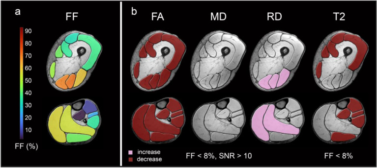 Figure 3
Peer Reviews
No public reviews on file for this paper yet. If you reviewed it on a platform where reviews are public (OpenReview, ICLR, NeurIPS, ICML), you can paste yours below so the community can read it here.
Videos
No videos yet. Explain this paper in a talk, walkthrough, or lecture? Add one.
Taxonomy
TopicsHistorical Economic and Social Studies
Correction to: Scientific Reports 10.1038/s41598-022-23972-6, published online 16 November 2022
The original version of this Article contained an error in Figure 3, where the colour code for ‘increase’ and ‘decrease’, stated within the image, was switched. The original Figure 3 and accompanying legend appear below.Figure 3qMRI data in low-fat muscles. Overview of mean fat fractions of all LGMD patients in thigh and calf muscles (a). High-risk muscles are coloured in yellow and orange, intermediate-risk muscles are coloured in green and low-risk muscles are coloured in blue. Muscle groups with significant differences of FA and MD in muscles with FF < 8% and SNR > 10 and T2 in muscle groups with FF < 8% between study groups are coloured in red (b) (increase/decrease in patient group: burgundy / pink).
The original Article has been corrected.