Use of High-density Mapping to Ablate a Wide Accessory Pathway and Mahaim Fiber in a Patient with Ebstein’s Anomaly
Eric Rosenthal, Ka-Chun Un, Julian Bostock

TL;DR
This paper describes using high-density mapping to successfully perform catheter ablation in a pediatric patient with Ebstein’s anomaly.
Contribution
The study is the first to demonstrate the utility of high-density mapping in ablation for Ebstein’s anomaly.
Findings
High-density mapping provided detailed anatomical and electrical insights in Ebstein’s anomaly.
The technique improved the success of catheter ablation in a challenging case.
It may reduce recurrence rates in similar patients.
Abstract
Ablation of accessory pathways in patients with Ebstein’s anomaly can be challenging. Despite increasing experience and advances in mapping technology, success is limited and recurrence rates can be high. To date, high-density electroanatomic mapping has not been studied in this anatomical substrate. We present a pediatric case of Ebstein’s anomaly in which high-density mapping in Ebstein’s anomaly was a useful additional tool to improve the outcome of catheter ablation.
Genes, proteins, chemicals, diseases, species, mutations and cell lines named across the full text — each resolved to its canonical identifier and authoritative record.
Click any figure to enlarge with its caption.
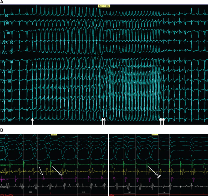 Figure 1
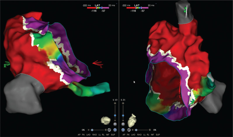 Figure 2
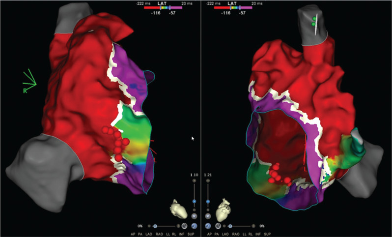 Figure 3
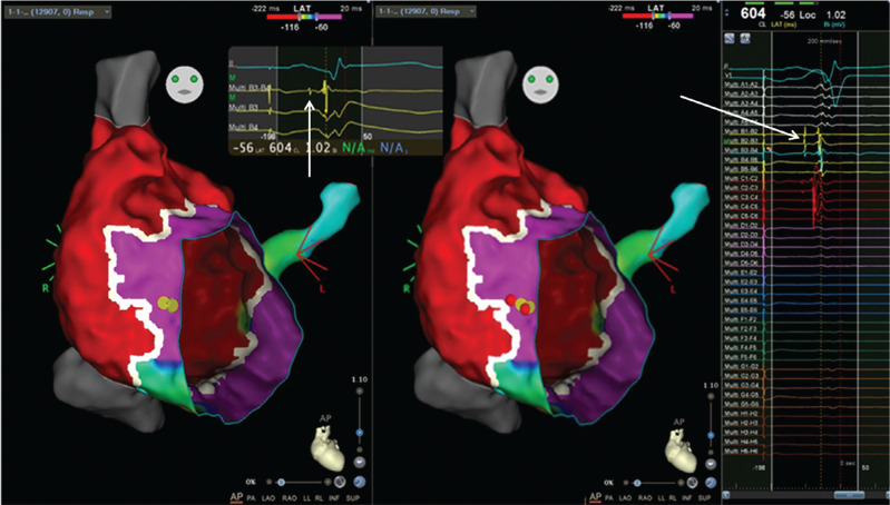 Figure 4
Peer Reviews
No public reviews on file for this paper yet. If you reviewed it on a platform where reviews are public (OpenReview, ICLR, NeurIPS, ICML), you can paste yours below so the community can read it here.
Videos
No videos yet. Explain this paper in a talk, walkthrough, or lecture? Add one.
Taxonomy
TopicsCardiac Arrhythmias and Treatments · Cardiovascular Issues in Pregnancy · Congenital Heart Disease Studies
Introduction
Ablation of accessory pathways in patients with Ebstein’s anomaly can be challenging due to the displacement of the anatomical annulus from the electrical annulus; low-amplitude signals over the atrialized portion of the right ventricle; and multiple accessory pathways, which can be broad. Despite increasing experience and advances in mapping technology, there is a lower success rate and a higher recurrence rate.^1–3^ High-density electroanatomic mapping has not been studied in this anatomical substrate. Additionally, in some patients, a Mahaim fiber coexists, adding to the complexity.^4,5^
Case presentation
An 11-year-old boy with Ebstein’s anomaly and moderate tricuspid valve displacement but only mild tricuspid regurgitation with overt pre-excitation developed supraventricular tachycardia and underwent a fluoroscopy-free electrophysiology study. During incremental atrial pacing, the accessory pathway became more overt, then changed to a pure left bundle branch block (LBBB) pattern with prolongation of the atrioventricular (AV) time before 2:1 AV block ensued with conduction across the AV node, revealing an underlying incomplete right bundle branch block (RBBB) pattern (Figures 1A and 1B). Atrial extrastimulus pacing induced runs of antidromic atrioventricular re-entrant tachycardia (AVRT) with an LBBB pattern and a long P–R interval. High-density mapping of the AV ring was performed during atrial pacing with maximum pre-excitation using an Octaray multipolar catheter with 2-mm spacing between electrodes on eight splines (Biosense Webster, Diamond Bar, CA, USA). Mapping was aided by a Vizigo sheath (Biosense Webster) in the difficult-to-reach areas. Open-window mapping (14,000 points) revealed a broad area of AV conduction on the posterior tricuspid annulus with continuous AV signals (Figure 2). Several ablations (Cool flow Navistar; Biosense Webster) in this area had a transient effect on accessory pathway conduction, but, after a linear row of lesions was delivered to the gap in the open-window map, there was no further conduction over the accessory pathway (Figure 3). Incremental atrial pacing now elicited an LBBB pattern that was not pre-excited, and antidromic AVRT was still inducible with the same pattern, suggestive of a Mahim fiber. A review of the high-density mapping electrograms revealed a “His-like” deflection at about 9 o’clock on the tricuspid annulus. The ablation catheter was unable to record a similar signal, even with amplification. Catheter pressure at the sites indicated by the electrograms on the open-window map resulted in the cessation of Mahaim conduction. Ablation was performed with no recurrence of Mahaim fiber conduction (Figure 4). The final ECG showed no pre-excitation with an incomplete RBBB pattern with decremental AV and ventriculoatrial conduction and non-inducibility of AVRT.
The patient’s parent provided written informed consent for case publication.
Discussion
Open-window mapping has recently been found to be useful in aiding ablation of conventional accessory AV pathways.^6–8^ In Ebstein’s anomaly, the AV annulus can be poorly defined due to the adherence of the tricuspid valve tissue to the ventricular myocardium, creating a zone of atrialized ventricular myocardium. This zone often has low-voltage signals that can be difficult to interpret and makes definition of the true electrical AV annulus difficult. The adherence of the tricuspid valve to the ventricular myocardium is also associated with multiple and broader accessory AV pathways. These features, together with poor catheter stability, can make ablation of accessory pathways in Ebstein’s anomaly challenging, with a lower success rate and a higher recurrence rate.^1–3^
High-density mapping with the open-window algorithm was initially beneficial both in identifying the electrical annulus and also demonstrating a broad area of accessory pathway conduction. Point ablations guided by signals were ineffective until a row of lesions was placed to “plug” the gap in the open-window map.
Mahaim fibers are also a feature of Ebstein’s syndrome, and mapping of these can be challenging, especially when the annulus is ill-defined.^4,5^ The Mahaim fiber signal on the normal AV annulus is not uncommonly indistinct when mapping with large surface area catheters, and these robust catheters may also block conduction inadvertently, preventing ablation. The high-density electrograms recorded with the delicate narrow-spaced electrodes of the Octaray catheter were able to identify the Mahaim potential clearly when the larger surface area electrodes of the ablation catheter were unable to detect it. This guided the ablation catheter to the site and allowed an effective ablation. High-density mapping has been used once before to demonstrate the potentials of a purely retrograde decremental accessory pathway in a concealed Mahaim-type structure on the right lateral AV groove in a 22-year-old man with hypertrophic cardiomyopathy and a normal tricuspid valve.^9^
Conclusions
High-density mapping in Ebstein’s anomaly may prove to be a useful additional tool to improve the outcome of catheter ablation in this anatomical substrate.
The reference list from the paper itself. Each links out to its DOI / PubMed record.
- 1El-Assaad I De Witt ES Mah DY Accessory pathway ablation in Ebstein anomaly: a challenging substrate Heart Rhythm 202118111844185110.1016/j.hrthm.2021.06.117134126268 · doi ↗ · pubmed ↗
- 2Karagöz T Ertuğrulİ Aypar E Two decades of experience on ablation in children with Ebstein’s anomaly Cardiol Young 202232343744310.1017/S 104795112100235334165064 · doi ↗ · pubmed ↗
- 3VukmirovićM Peichl P Kautzner J Catheter ablation of multiple accessory pathways in Ebstein anomaly guided by intracardiac echocardiography Europace 201618333910.1093/europace/euv 42826851810 · doi ↗ · pubmed ↗
- 4Ueshima K Nakamura Y Takeno S Miyake T Takemura T Atriofascicular Mahaim with Ebstein anomaly: a case report J Arrhythm 201733550851010.1016/j.joa.2017.06.00729021860 PMC 5634721 · doi ↗ · pubmed ↗
- 5Viray MC Wiener PC Batnyam U Rasquin L Pressman GS Mainigi S A young woman with recurrent palpitations: a case of Ebstein anomaly with Mahaim fiber tachycardia CASE (Phila)20193414514810.1016/j.case.2019.03.00631468016 PMC 6710817 · doi ↗ · pubmed ↗
- 6Frisch DR High-density mapping of a posteroseptal accessory pathway using open-window mapping J Innov Cardiac Rhythm Manage 202112 S 1111310.19102/icrm.2021.120102 S 33604106 PMC 7885966 · doi ↗ · pubmed ↗
- 7Schricker AA Winkle R Moskovitz R Open-window mapping of accessory pathways utilizing high-density mapping J Interv Card Electrophysiol 202161352553310.1007/s 10840-020-00850-732789708 · doi ↗ · pubmed ↗
- 8Regan W Harris K Rosenthal E Opening a window into accessory pathway mapping in children with Wolff-Parkinson-White Syndrome?J Innov Card Rhythm Manag 202213115222522410.19102/icrm.2022.1311436570483 PMC 9721299 · doi ↗ · pubmed ↗