Exploration of neuroanatomical characteristics to differentiate prodromal Alzheimer’s disease from cognitively unimpaired amyloid-positive individuals
Hak Hyeon Kim, Min Jeong Kwon, Sungman Jo, Ji Eun Park, Ji Won Kim, Jae Hyoung Kim, Sang Eun Kim, Ki Woong Kim, Ji Won Han

TL;DR
This study identifies brain regions where volume loss, not amyloid buildup, distinguishes early Alzheimer’s from amyloid-positive but cognitively normal individuals.
Contribution
The study introduces a novel approach combining amyloid PET and MRI to identify neuroanatomical markers specific to prodromal Alzheimer’s disease.
Findings
Regional volume differences in the amygdala, hippocampus, and entorhinal cortex distinguish prodromal AD from cognitively unimpaired amyloid-positive individuals.
The interaction between amyloid deposition and reduced volume in the superior parietal cortex increases the likelihood of prodromal AD.
Amyloid deposition levels alone do not differentiate prodromal AD from cognitively unimpaired amyloid-positive individuals.
Abstract
Differentiating clinical stages based solely on positive findings from amyloid PET is challenging. We aimed to investigate the neuroanatomical characteristics at the whole-brain level that differentiate prodromal Alzheimer’s disease (AD) from cognitively unimpaired amyloid-positive individuals (CU A+) in relation to amyloid deposition and regional atrophy. We included 45 CU A+ participants and 135 participants with amyloid-positive prodromal AD matched 1:3 by age, sex, and education. All participants underwent 18F-florbetaben positron emission tomography and 3D structural T1-weighted magnetic resonance imaging. We compared the standardized uptake value ratios (SUVRs) and volumes in 80 regions of interest (ROIs) between CU A+ and prodromal AD groups using independent t-tests, and employed the least absolute selection and shrinkage operator (LASSO) logistic regression model to identify…
Genes, proteins, chemicals, diseases, species, mutations and cell lines named across the full text — each resolved to its canonical identifier and authoritative record.
Click any figure to enlarge with its caption.
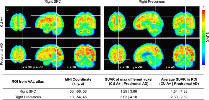 Figure 1
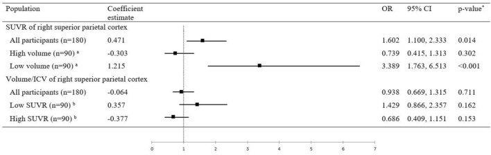 Figure 2
Peer Reviews
No public reviews on file for this paper yet. If you reviewed it on a platform where reviews are public (OpenReview, ICLR, NeurIPS, ICML), you can paste yours below so the community can read it here.
Videos
No videos yet. Explain this paper in a talk, walkthrough, or lecture? Add one.
Taxonomy
TopicsDementia and Cognitive Impairment Research · Alzheimer's disease research and treatments · Functional Brain Connectivity Studies