Interest of Chest CT to Assess the Prognosis of SARS-CoV-2 Pneumonia: An In-Hospital-Based Experience in Sub-Saharan Africa
Serge Emmanuel Obe -A- Ndzem Holenn, Tacite Kpanya Mazoba, Désiré Yaya Mukanga, Tyna Bongosepe Zokere, Djo Lungela, Jean-Robert Makulo, Steve Ahuka, Angèle Tanzia Mbongo, Antoine Aundu Molua

TL;DR
This study shows that chest CT scans can help predict the risk of death in patients with suspected SARS-CoV-2 pneumonia in Sub-Saharan Africa.
Contribution
The study demonstrates that lung lesion extent on chest CT is a strong predictor of mortality in confirmed and suspected SARS-CoV-2 pneumonia cases.
Findings
Chest CT features like ground glass and consolidation were more common in confirmed SARS-CoV-2 cases.
Lung lesions affecting 30% of the parenchyma predicted mortality with moderate accuracy (AUC 0.705).
Lesions affecting 50% of the lung parenchyma increased the risk of death by 7.194 times.
Abstract
We included all patients with respiratory symptoms (dyspnea, fever, and cough) and/or respiratory failure admitted to the SOS Médecins de nuit SARL hospital, DR Congo, during the 2nd and 3rd waves of the COVID-19 pandemic. The diagnosis of COVID-19 was established based on RT-PCR anti-SARS-CoV-2 tests (G1 (RT-PCR positive) vs. G2 (RT-PCR negative)), and all patients had a chest CT on the day of admission. We retrieved the digital files of patients, precisely the clinical, biological, and chest CT parameters of the day of admission as well as the vital outcome (survival or death). Chest CT were read by a very high-definition console using Advantage Windows software and exported to the hospital network using the RadiAnt DICOM viewer. To determine the threshold for the percentage of lung lesions associated with all-cause mortality, we used ROC curves. Factors associated with death,…
Genes, proteins, chemicals, diseases, species, mutations and cell lines named across the full text — each resolved to its canonical identifier and authoritative record.
Click any figure to enlarge with its caption.
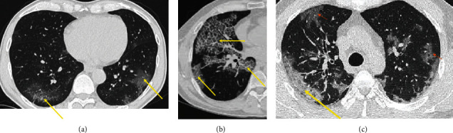 Figure 1
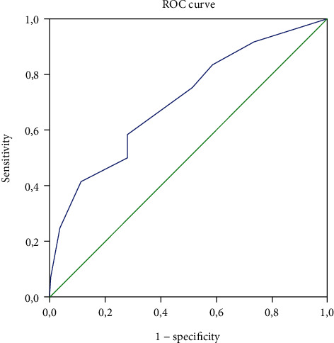 Figure 2
Peer Reviews
No public reviews on file for this paper yet. If you reviewed it on a platform where reviews are public (OpenReview, ICLR, NeurIPS, ICML), you can paste yours below so the community can read it here.
Videos
No videos yet. Explain this paper in a talk, walkthrough, or lecture? Add one.
Taxonomy
TopicsCOVID-19 diagnosis using AI · COVID-19 Clinical Research Studies · Ultrasound in Clinical Applications