Computational Approaches and Observer Variation in the 3D Musculoskeletal Modeling of the Heads of Anolis
A D Lagorio, F R McGechie, M G Fields, J Fortner, E Mackereth, C Perez, A T Wilken, M Leal, C V Ward, K M Middleton, C M Holliday
TL;DR
This paper explores new 3D modeling techniques to study the musculoskeletal anatomy of two Anolis lizard species, highlighting the repeatability and potential of these methods for evolutionary research.
Contribution
The study introduces and evaluates novel 3D modeling approaches for quantifying musculoskeletal variation in Anolis lizards, including muscle segmentation and attachment mapping.
Findings
3D modeling techniques like DiceCT segmentation and surface attachment mapping are repeatable and accessible for musculoskeletal analysis.
The study provides new data on jaw muscle volumes and fiber architecture in A. pulchellus and A. sagrei.
Sexual dimorphism in skull anatomy is observed, offering insights into evolutionary and biomechanical adaptations.
Abstract
High-resolution imaging, 3D modeling, and quantitative analyses are equipping evolutionary biologists with new approaches to understanding the variation and evolution of the musculoskeletal system. However, challenges with interpreting DiceCT data and higher order use of modeled muscles have not yet been fully explored, and the error in and accuracy of some digital methods remain unclear. West Indian Anolis lizards are a model clade for exploring patterns in functional adaptation, ecomorphology, and sexual size dimorphism in vertebrates. These lizards possess numerous jaw muscles with potentially different anatomies that sculpt the adductor chamber of the skull. Here we test approaches to quantifying the musculoskeletal shape of the heads of two species of Anolis: A. pulchellus and A. sagrei. We employ comparative approaches such as DiceCT segmentation of jaw muscles, 3D surface…
Genes, proteins, chemicals, diseases, species, mutations and cell lines named across the full text — each resolved to its canonical identifier and authoritative record.
Click any figure to enlarge with its caption.
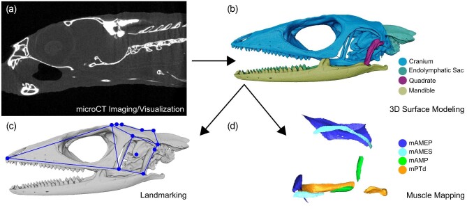 Figure 1
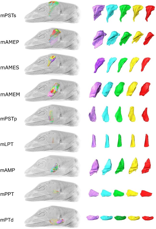 Figure 10
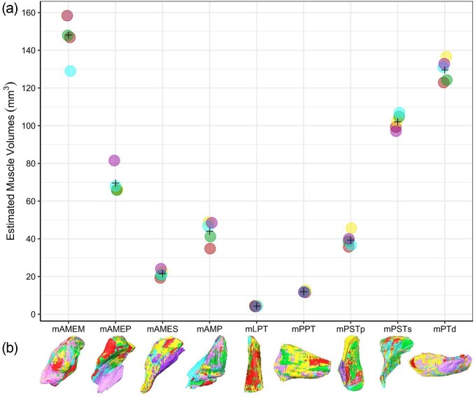 Figure 11
 Figure 12
 Figure 13
 Figure 2
 Figure 3
 Figure 4
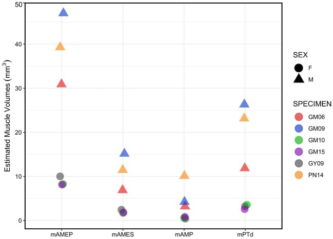 Figure 5
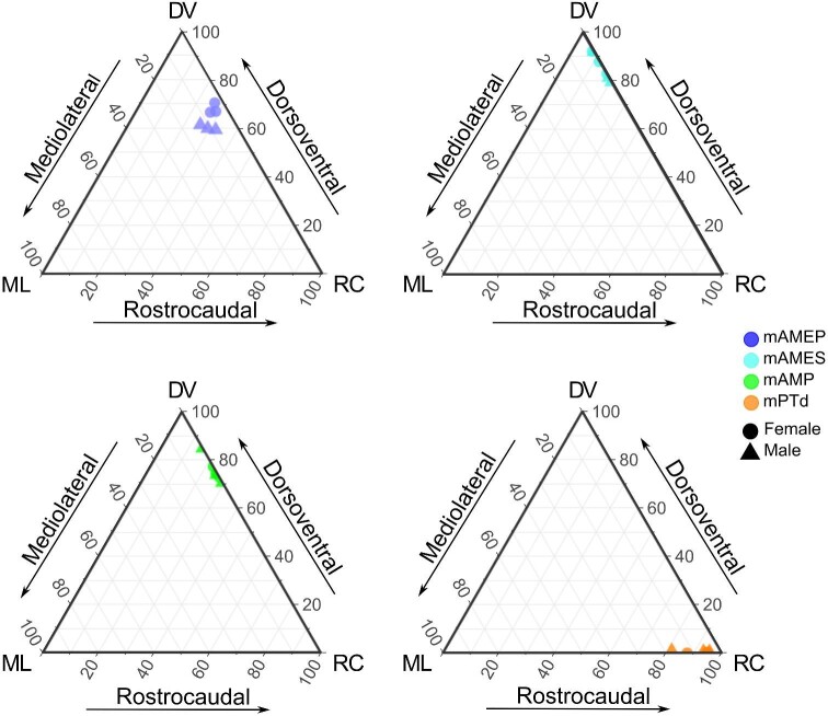 Figure 6
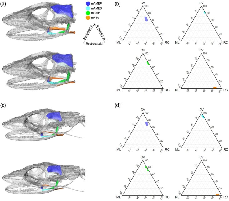 Figure 7
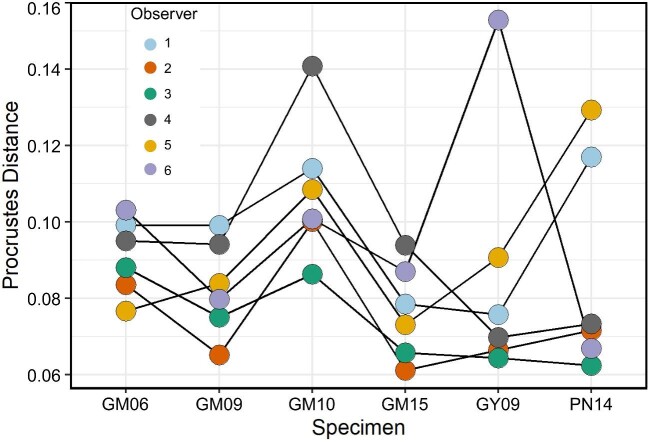 Figure 8
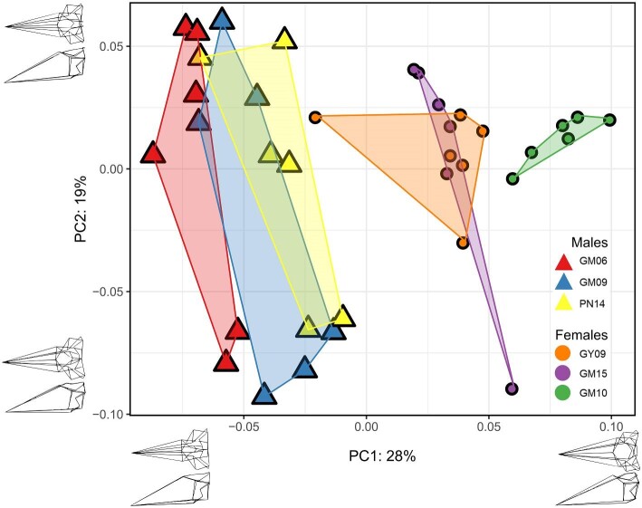 Figure 9
Peer Reviews
No public reviews on file for this paper yet. If you reviewed it on a platform where reviews are public (OpenReview, ICLR, NeurIPS, ICML), you can paste yours below so the community can read it here.
Videos
No videos yet. Explain this paper in a talk, walkthrough, or lecture? Add one.
Taxonomy
TopicsMorphological variations and asymmetry