The prone position in COVID-19 impacts the thickness of peripapillary retinal nerve fiber layers and macular ganglion cell layers
Niphon Chirapapaisan, Akarawit Eiamsamarng, Wanicha Chuenkongkaew, Natthapon Rattanathamsakul, Ranistha Ratanarat, Karim Adly Raafat, Karim Adly Raafat

TL;DR
Placing COVID-19 patients in the prone position affects retinal layers, but does not increase eye-related health risks.
Contribution
This study is the first to investigate how the prone position affects retinal nerve fiber and ganglion cell layers in COVID-19 patients.
Findings
The prone position in COVID-19 patients leads to early loss of peripapillary retinal nerve fiber layer thickness.
No significant differences in retinal nerve fiber layer thickness were found between prone and non-prone groups at three follow-ups.
The macular ganglion cell layer was thinner in non-prone patients compared to controls at 1 and 6 months.
Abstract
The prone position reduces mortality in severe cases of COVID-19 with acute respiratory distress syndrome. However, visual loss and changes to the peripapillary retinal nerve fiber layer (p-RNFL) and the macular ganglion cell layer and inner plexiform layer (m-GCIPL) have occurred in patients undergoing surgery in the prone position. Moreover, COVID-19-related eye problems have been reported. This study compared the p-RNFL and m-GCIPL thicknesses of COVID-19 patients who were placed in the prone position with patients who were not. This prospective longitudinal and case-control study investigated 15 COVID-19 patients placed in the prone position (the “Prone Group”), 23 COVID-19 patients not in the prone position (the “Non-Prone Group”), and 23 healthy, non-COVID individuals without ocular disease or systemic conditions (the “Control Group”). The p-RNFL and m-GCIPL thicknesses of the…
Genes, proteins, chemicals, diseases, species, mutations and cell lines named across the full text — each resolved to its canonical identifier and authoritative record.
Click any figure to enlarge with its caption.
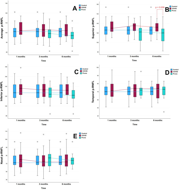 Figure 1
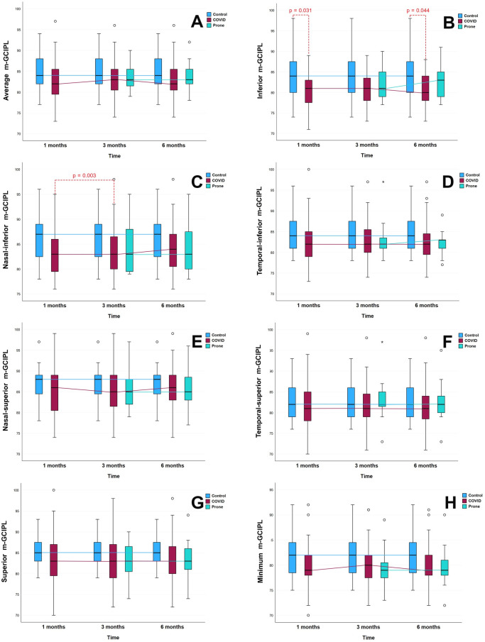 Figure 2
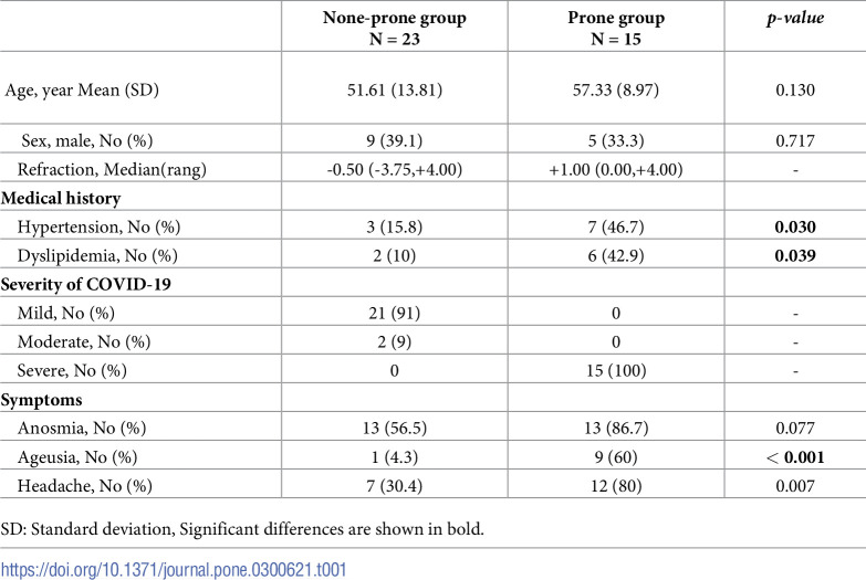 Figure 3
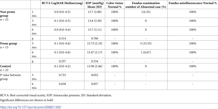 Figure 4
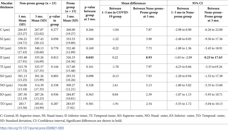 Figure 5
Peer Reviews
No public reviews on file for this paper yet. If you reviewed it on a platform where reviews are public (OpenReview, ICLR, NeurIPS, ICML), you can paste yours below so the community can read it here.
Videos
No videos yet. Explain this paper in a talk, walkthrough, or lecture? Add one.
Taxonomy
TopicsRetinal and Optic Conditions · Intraoperative Neuromonitoring and Anesthetic Effects · Glaucoma and retinal disorders