Hepatic Duct Adenoma Identified Using Direct Cholangioscopy
Butros Fakhoury, Iyiad Alabdulrazzak, Michael Talanian, Syed Mahmood

TL;DR
A rare bile duct tumor was diagnosed in a 46-year-old woman using direct cholangioscopy, emphasizing its value in identifying such lesions.
Contribution
Demonstrates the utility of direct cholangioscopy in diagnosing bile duct adenomas.
Findings
Direct cholangioscopy identified a 15 mm polypoid lesion in the common hepatic duct.
Biopsy confirmed the lesion as a bile duct adenoma.
Abstract
Bile duct adenomas (BDAs) are rare benign tumors that can arise in the intra-hepatic or extra-hepatic biliary tree. We present a case of a 46-year-old female who presented with symptoms suggestive of choledocholithiasis. Direct cholangioscopy identified a 15 mm polypoid lesion in the common hepatic duct (CHD). Biopsy revealed a BDA. We present this case to highlight the role of direct cholangioscopy in the diagnosis and management of BDAs.
Genes, proteins, chemicals, diseases, species, mutations and cell lines named across the full text — each resolved to its canonical identifier and authoritative record.
Click any figure to enlarge with its caption.
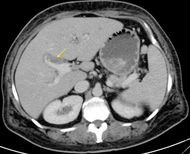 Figure 1
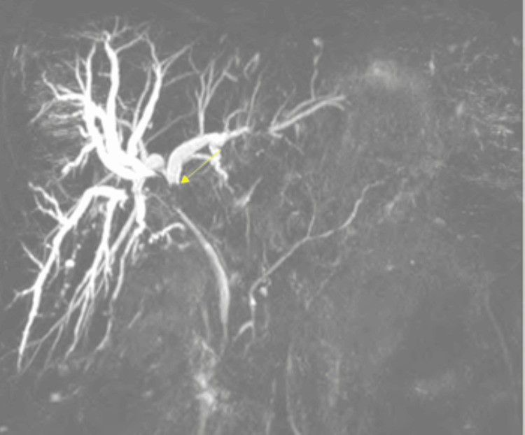 Figure 2
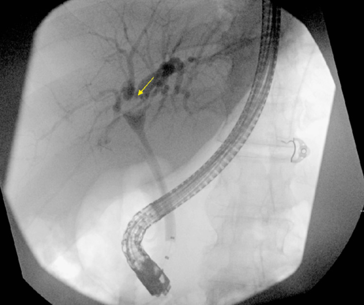 Figure 3
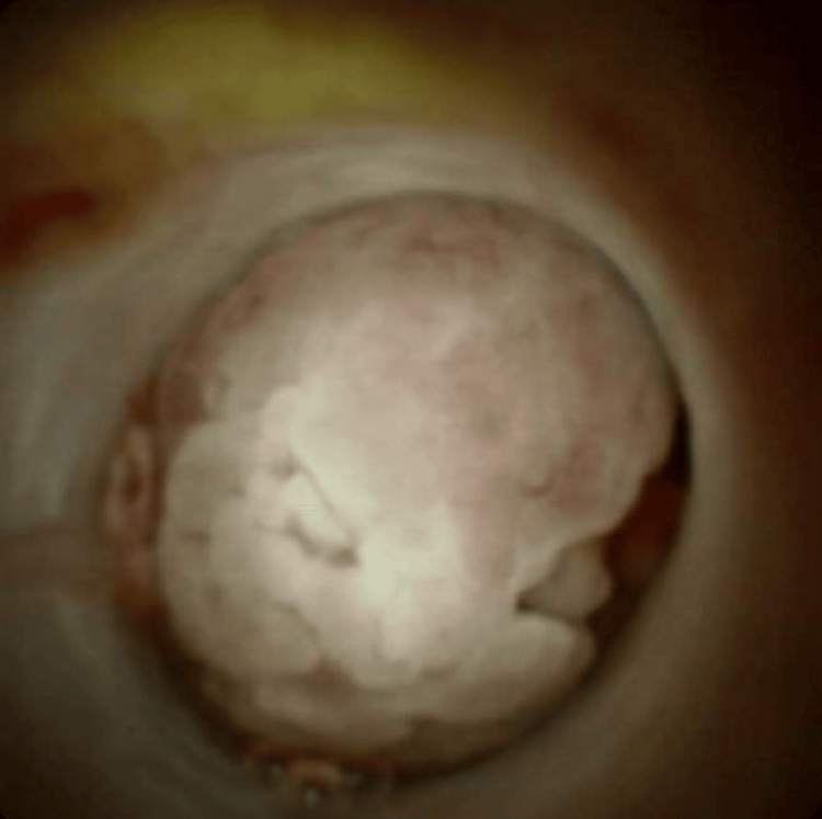 Figure 4
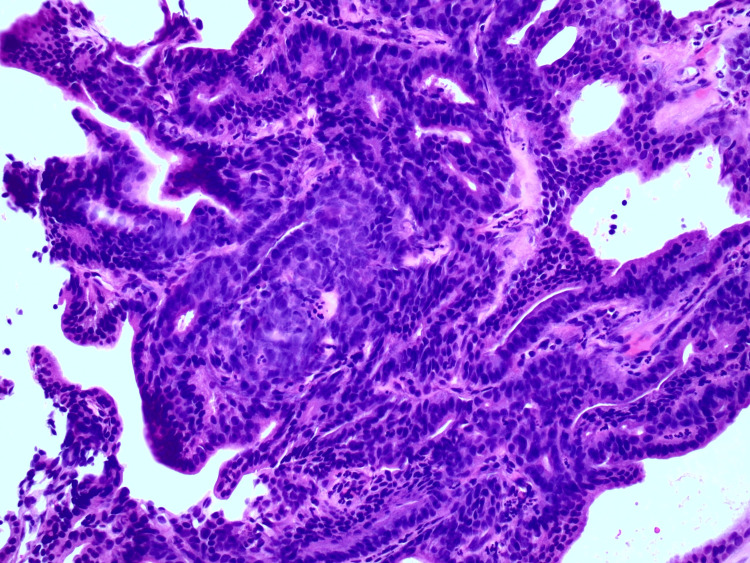 Figure 5
Peer Reviews
No public reviews on file for this paper yet. If you reviewed it on a platform where reviews are public (OpenReview, ICLR, NeurIPS, ICML), you can paste yours below so the community can read it here.
Videos
No videos yet. Explain this paper in a talk, walkthrough, or lecture? Add one.
Taxonomy
TopicsCholangiocarcinoma and Gallbladder Cancer Studies · Genetic and Kidney Cyst Diseases · Pediatric Hepatobiliary Diseases and Treatments