Awareness for artifacts in fluorescence microscopy of β-TCP
Marco Waldmann, Marc Bohner, Anna Baghnavi, Bianca Riedel, Michael Seidenstuecker

TL;DR
This paper highlights previously unknown artifacts in fluorescence microscopy of β-TCP ceramics that can be mistaken for cells and suggests ways to reduce them.
Contribution
The paper identifies and proposes solutions for new artifacts in β-TCP fluorescence microscopy that may lead to false positives.
Findings
Artifacts in β-TCP fluorescence microscopy can resemble cells in shape and fluorescence behavior.
Technovit 9100 fixation and mechanical cleaning significantly reduce these artifacts.
Untreated dowels showed no reduction in artifacts regardless of further treatment.
Abstract
Fluorescence analysis of β-TCP ceramics is often used to describe cells found on said ceramics. However, we found, to our knowledge, so far undescribed artifacts which might sometimes be hard to differentiate from cells due to shape and fluorescence behavior. We tried prolonged ultrasound washing as well as Technovit 9100 fixation to reduce these artifacts. While untreated dowels showed no reduction in artifacts no matter the further treatment, Technovit fixation reduced the artifacts with even further reduction achieved by mechanical cleaning. As a consequence, scientists working with these dowels and likely even other types should try to avoid creating false positive results by considering the existence of these artifacts, checking additional filters for unusual fluorescence and by reducing them by using Technovit fixation when possible.
Genes, proteins, chemicals, diseases, species, mutations and cell lines named across the full text — each resolved to its canonical identifier and authoritative record.
Click any figure to enlarge with its caption.
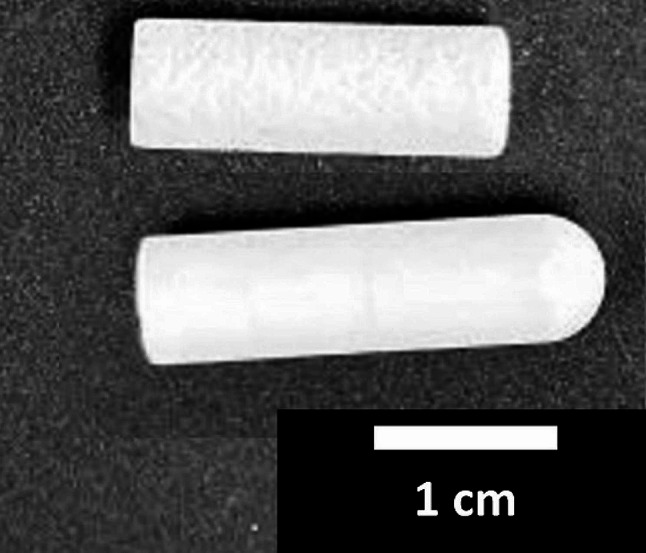 Figure 1
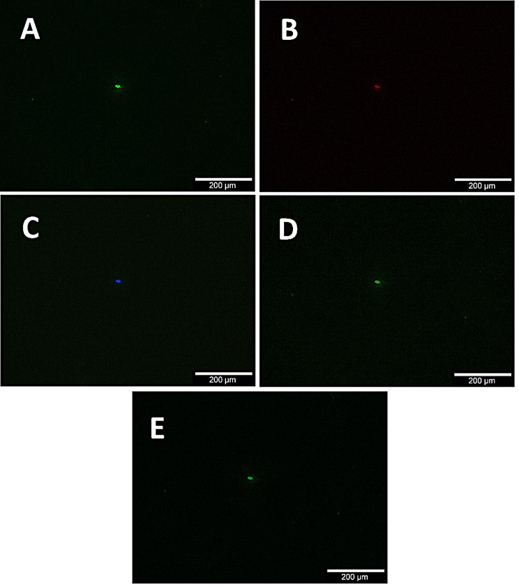 Figure 2
- —Universitätsklinikum Freiburg (8975)
Peer Reviews
No public reviews on file for this paper yet. If you reviewed it on a platform where reviews are public (OpenReview, ICLR, NeurIPS, ICML), you can paste yours below so the community can read it here.
Videos
No videos yet. Explain this paper in a talk, walkthrough, or lecture? Add one.
Taxonomy
TopicsPhotoacoustic and Ultrasonic Imaging · Thermography and Photoacoustic Techniques · Ultrasonics and Acoustic Wave Propagation
Introduction
β-TCP ceramics are frequently used in orthopedic settings and well researched [1–6]. They are biodegradable ceramics which hold osteoinductive and osteoconductive properties and are able to form a strong bond with host tissue. The mechanical properties of β-TCP however are limited. They can be seen as an alternative to the “gold standard” of autologous bone grafts, which, although they have high osteoinductive and osteogenic capacities, come with disadvantages like an increased risk of infection due to a second surgical site, limitation of available graft material and likewise limited mechanical properties, making them a great choice for smaller but not for large-scale bone defects. In situations where these disadvantages outweigh the advantages of autologous bone grafts, alternatives like β-TCP come into play [7–9].
Research of β-TCP includes analysis of surfaces using fluorescence microscopy as one common technique to characterize cells found on these surfaces. For example, live/dead assays to access viability of cells [10], immunofluorescence [10] or proof of loading using FITC conjugated drugs [11] are common practice. Nonetheless, fluorescence microscopy can also lead to unwanted effects or artifacts, possibly resulting in misinterpretation. The process of photobleaching describes the loss of fluorescence of a fluorochrome molecule due to chemical destruction after exposure to excitation light [12]. Consequently, originally positive signals might be missed if the excitation time is too long. Techniques to examine specific proteins like immunolabeling also rely on the specificity of the primary antibody in use [13]. Without proper testing, unintended proteins might also produce positive signals. With immunolabeling, incomplete protein tagging can also occur if the targeted cells are not be properly permeabilized [13].
Autofluorescence describes the inherent fluorescence without addition of an exogenous fluorescent agent found in some substances [14]. This phenomenon can be found in various instances. Examples include certain plastics [15], plants [16] and also human tissue [17, 18]. Depending on the signal strength, this phenomenon could also be falsely interpreted as a fluorescence caused by addition of external fluorescent molecules, if not properly accounted for in the first place. Another problem might occur, when different fluorescent agents with overlapping wavelength ranges of emitted light are chosen and not properly separated with adequate filters [19].
During our analysis of β-TCP, we noticed artifacts that, to our knowledge, have not yet been addressed and could potentially be confused with cells during fluorescence microscopy in an appropriate context.
Material
MaterialInformationEthanol 99%SAV Liquid Production GmBH, Flintsbach am Inn, GermanyTechnovit 9100 Combipack (contains: 1 × 1000 ml basic solution 1 × 120 g PMMA powder 8 × 1 g hardener 1 1 × 10 ml hardener 2 1 × 5 ml regulator), #64,715,444Kulzer GmbH, Hanau, GermanyCerasorb M Cylinder (Lot D147.501043)Curasan, Kleinostheim, GermanyKimtech® Science Precision Wipes, #7552Irving, Tx, USAFilter: Alizarin/Xylenolorange; Calcein; Cy5&AF647; Tetracyclin; FITC/Cy5 H Dualband FilterAHF, Tuebingen, GermanyOlympus BX51 fluorescent microscope, equipped with a 10x objectiveOlympus, Tokyo, JapanUV-Lightsource X-Cite Series 120 QExcelitas Technologies, Waltham, USA
Methods
We analyzed the surface of microporous β-TCP dowels (Ø 7 mm x L 26 mm, median pore diameter 5 μm, 40% total porosity [20], Fig. 1) produced by the RMS according to our specifications [21, 22] using fluorescence microscopy. To reduce the artifacts we tried different procedures of either ultrasound bathing dowels in 70% ethanol followed by distilled water for 10 min each (frequency 80/50 Hz, 26 °C) or ultrasound bathing in 70% ethanol followed by distilled water for 2 h with the same parameters. Sucking 5 mL of distilled water through the ceramics by applying a slight vacuum (650 mBar) to a flow chamber [23, 24] was tested as another method of reducing the artifacts. After ultrasound bathing for 10 min each, some dowels were fixated using Technovit 9100 new. Mechanical cleaning using cellulose wipes was tried in addition for Technovit fixated dowels and dowels ultrasound bathed for 10 min each. Untreated dowels (Ø 7 mm x L 20 mm, median pore diameter 37 μm, 62% total porosity [20], Fig. 1) purchased from a different manufacturer, Curasan (Cersasorb M, Fig. 1), were also analyzed.
Images of the dowels were taken at a defined position using an Olympus BX51 fluorescent microscope equipped with a 10x objective (Olympus, Tokyo, Japan) and filters for five different wavelengths. Artifact counts were determined for the whole image using ImageJ automated particle counting for images captured with a FITC/Cy5 H Dualband filter. All data are presented as mean ± standard deviation.
Results
The artifacts varied in size and shape but were often found to be round and had diameters ranging from approximately 5 to 80 μm. The artifacts were found on every dowel regardless of procedure and were fluorescent for all five filters analyzed (Fig. 2). Completely untreated (8.67 ± 0.58), ultrasound bathed for 10 min each (12 ± 7.21) as well as dowels ultrasound bathed for 2 h each (8.67 ± 5.03) showed said shapes for all wavelengths analyzed with no reduction in any group. Dowels fixated in Technovit showed a reduced number of artifacts (3.67 ± 0.58) compared to untreated dowels. For Technovit fixated dowels, mechanical cleaning reduced the artifacts even further (1.33 ± 0.58) but despite all efforts never achieved complete removal. Mechanical cleaning showed no effect for dowels ultrasound bathed for 10 min each (12.00 ± 1.73). (Table 1)
Untreated dowels by Curasan, were found to show said artifacts with slightly lower numbers than RMS dowels (5.33 ± 1.15). Cleaning the ceramics using 5 mL of distilled water under a slight vacuum was unsuccessful in reducing the artifacts (7.33 ± 2.31). (Table 1)
Fig. 1. Picture of the two types of dowels analyzed within this work. The upper dowel was manufactured by Curasan, the lower dowel was produced by the RMS. A scalebar in cm is provided within the picture
Table 1. Result of ImageJ counting for images captured with a FITC/Cy5 H Dualband filter of each analyzed group (n = 3) presented as mean ± standard deviationAnalyzed dowelsArtifactsCurasanuncleaned5.33 ± 1.15RMSuncleaned8.67 ± 0.5810 min ultrasound bath12 ± 7.2110 min ultrasound bath, mechanically cleaned12 ± 1.732 h ultrasound bath8.67 ± 5.03slight vacuum7.33 ± 2.31Technovit3.67 ± 0.58Technovit, mechanically cleaned1.33 ± 0.58
Fig. 2. Exemplary picture of fluorescence artifacts found on untreated, microporous β-TCP dowels provided by the RMS. All Frames show the same area using different excitation filters for Alizarin/Xylenolorange (Frame A), Tetracyclin (Frame B), Cy5&AF647 (Frame C), FITC/Cy5 H Dualband Filter (Frame D) and Calcein (Frame E). The Pictures were taken with an Olympus BX51 fluorescent microscope equipped with a 10x objective (Olympus, Tokyo, Japan)
Discussion
While the origin of the observed artifacts is unknown to us, impurities during the production process or surface modification during ceramic sintering [25] seem to be a reasonable explanation. The artifacts may be limited to the analyzed type of β-TCP samples produced by RMS and Curasan but for the evaluation of cells on β-TCP by fluorescence microscopy, researchers should be aware of the artifacts described in this note and prevent false positive results by considering their existence, even for other β-TCP products. With no fluorescence filter producing negative results, the artifacts can also be expected to extent to different wavelengths.
In general, in regard of the described artifacts, Technovit fixation followed by cautious mechanical cleaning seems to be the most suitable procedure, if tolerated by the type of sample that has to be evaluated. In addition, and especially when untreated dowels are analyzed, checking a completely untreated dowel for the described unwanted artifacts as a reference, as well as separating artifacts from actual positive results by controlling other wavelengths that would not normally show fluorescence signals with the chosen parameters, seems to be a suitable method for preventing false positive results. Ultrasound bathing probably does not help to reduce this type of artifact.
Another possible option to reduce the effect of autofluorescence is the exploitation of differences in fluorescence lifetimes between autofluorescent substances and fluorescent agents with longer lifetimes [26–28].
Assuming autofluorescence of impurities during production as the source of artifacts and depending on the fluorescent agents in use, trying to find differences between the artifacts and structures labeled with exogenous markers via means of fluorescence lifetime imaging microscopy [29, 30] may prove useful in the future to correctly differentiate these artifacts from cells.
Limitations
Only dowels produced by the RMS and Curasan, and only from one batch each, were analyzed. Also, the filters were limited to five different types, while we expect the bandwidth of fluorescenting wavelengths to be even broader than observed in this work.
The reference list from the paper itself. Each links out to its DOI / PubMed record.
- 1Agarwalla A Puzzitiello R Garcia GH Forsythe B Application of a Beta-tricalcium phosphate graft to Minimize Bony defect in Bone-Patella Tendon-Bone Anterior Cruciate Ligament Reconstruction Arthrosc Tech 201877 e 725910.1016/j.eats.2018.03.00930094143 PMC 6074020 · doi ↗ · pubmed ↗
- 2Cho CH Bae KC Kim DH Biomaterials used for suture anchors in orthopedic surgery Clin Orthop Surg 20211332879210.4055/cios 2031734484619 PMC 8380519 · doi ↗ · pubmed ↗
- 3Jeong JH Jin ES Kim JY Lee B Min J Jeon SR The Effect of Biocomposite screws on Bone Regeneration in a rat osteoporosis model World Neurosurg 20171069647210.1016/j.wneu.2017.07.08328736352 · doi ↗ · pubmed ↗
- 4Tai S Cheng JY Ishii H Akimoto S Satoh T Yamamoto K Characterization of beta-tricalcium phosphate as a novel immunomodulator Int Immunopharmacol 2014191455110.1016/j.intimp.2013.12.02424412469 · doi ↗ · pubmed ↗
- 5Dorozhkin SV Amorphous calcium (Ortho)phosphates Acta Biomater 201061244577510.1016/j.actbio.2010.06.03120609395 · doi ↗ · pubmed ↗
- 6Dorozhkin SV Calcium orthophosphates as Bioceramics: state of the art J Funct Biomaterials 2010112210710.3390/jfb 1010022 PMC 403089424955932 · doi ↗ · pubmed ↗
- 7Li X Zhang H Zhang H Fabrication of β-TCP/ akermanite composite scaffold via DLP and in-situ modification of micro-nano surface morphology for bone repair Ceram Int 202450226596910.1016/j.ceramint.2023.10.276 · doi ↗
- 8Gillman CE Jayasuriya ACFDA-approved bone grafts and bone graft substitute devices in bone regeneration Mater Sci Engineering: C 202113011246610.1016/j.msec.2021.112466 PMC 855570234702541 · doi ↗ · pubmed ↗