Baseline CD4+ and expansion of γδ T cells correlate with response to durvalumab in triple‐negative breast cancer patients
Chiara Massa, Thomas Karn, Karsten Weber, Andreas Schneeweiss, Claus Hanusch, Jens Uwe Blohmer, Dirk‐Michael Zahm, Christian Jackisch, Marion van Mackelenbergh, Jörg Thomalla, Frederik Marmé, Jens Huober, Volkmar Müller, Christian Schem, Anja Müller, Elmar Stickeler

Abstract
Genes, proteins, chemicals, diseases, species, mutations and cell lines named across the full text — each resolved to its canonical identifier and authoritative record.
Click any figure to enlarge with its caption.
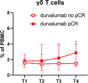 Figure 1
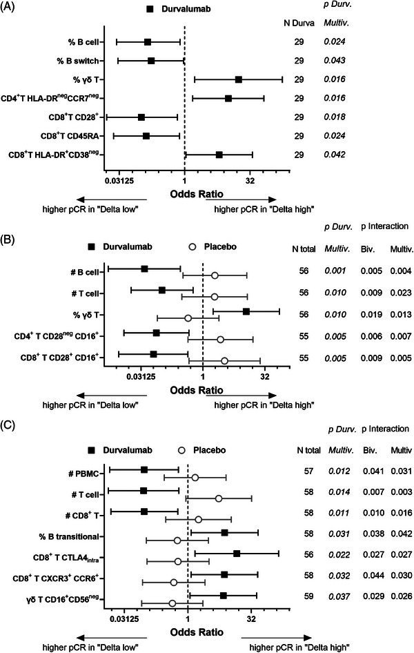 Figure 2
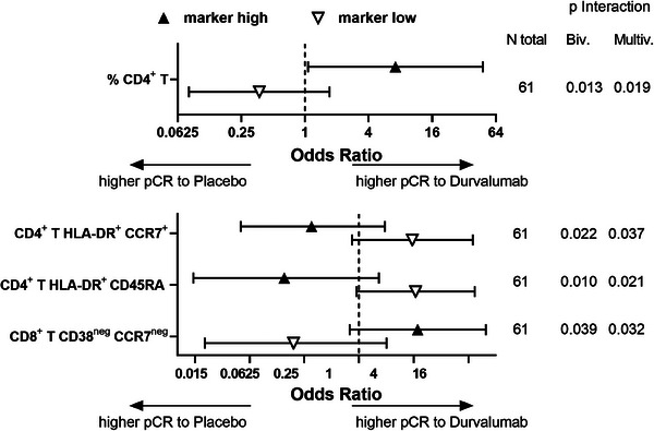 Figure 3
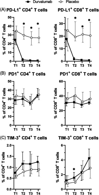 Figure 4
- —Celgene 10.13039/100006436
- —German Cancer Aid 10.13039/501100005972
Peer Reviews
No public reviews on file for this paper yet. If you reviewed it on a platform where reviews are public (OpenReview, ICLR, NeurIPS, ICML), you can paste yours below so the community can read it here.
Videos
No videos yet. Explain this paper in a talk, walkthrough, or lecture? Add one.
Taxonomy
TopicsCancer Immunotherapy and Biomarkers · CAR-T cell therapy research · Immunotherapy and Immune Responses
Dear Editor,
Immunomonitoring of patients with primary, non‐metastatic triple‐negative breast cancer (TNBC) from the GeparNuevo trial indicated that treatment with the immune checkpoint inhibitor (ICPi) durvalumab resulted in almost complete coverage of its target programmed death ligand 1 (PD‐L1) on circulating immune cells. Moreover, pathological complete response (pCR) upon the addition of durvalumab to neoadjuvant chemotherapy (NAC) correlated with higher pretreatment levels of CD4^+^ T cells and with expansion of γδ T cells during treatment.
Since TNBC lacks targetable disease drivers, but has a high lymphocytic infiltration, different attempts to implement immunotherapeutic approaches have been performed including ICPi, which improved clinical responses only in a limited number of patients thereby underlying the need to identify predictive biomarkers to better stratify patients for therapy.1
In this study, 63 TNBC patients of the window sub‐cohort of the randomized, double‐blind phase II GeparNuevo trial treated with NAC in the presence or absence of the anti‐PD‐L1 Ab durvalumab2 were analyzed. Blood was drawn at four different time points [i.e. T1 at baseline, T2 after the window treatment with durvalumab, T3 after nanoparticle‐bound paclitaxel (Nab‐Pac) and T4 at surgery after treatment with epirubicin and cyclophosphamide (EC); Figure S1] and characterized by multicolour flow cytometry using different Ab panels (Table S1). The baseline characteristics of TNBC patients undergoing immunomonitoring were not statistically different from those of the overall study (Table S2). However, the increase in pCR to durvalumab versus placebo in patients of the window sub‐cohort reached significance in the total trial,2 but not in the immunomonitored patients (64.7% vs. 58.6%, respectively, odds ratio [OR] = 1.29, 95% confidence interval 0.47–3.59, p = .620; Figure S2).
Characterization of the immune cell absolute counts, frequencies and phenotype during treatment highlighted many alterations (Table S3). The most consistent was the almost complete loss of PD‐L1 molecules on circulating cells already after the first application (Figure 1A). Since the Ab moiety of durvalumab has been engineered to avoid interaction with the complement and Fc receptors,3 this reduced detection is due to shielding of PD‐L1 and not due to the elimination of PD‐L1‐positive cells, as confirmed by an equal absolute number and frequency of T cells in durvalumab‐ and placebo‐treated patients (Figure S3).
The only marginally more frequent pCR to durvalumab despite the homogenous loss of PD‐L1 availability might be due to different reasons. Despite NAC caused an upregulation of PD1 and TIM‐3 on T cells in the placebo arm of GeparNuevo,4 only a transient upregulation of TIM‐3 on CD8^+^ T cells was found after the window treatment with durvalumab (Figure 1B,C) thereby excluding the involvement of additional immune escape mechanisms in the failure to enhance pCR.5 An alternative explanation might be that the loss of the availability of PD‐L1 within the tumours is not as complete as in the periphery, as demonstrated in a murine setting.6
The search of predictive immune cell marker(s) for patients´ stratification indicated that high frequencies of CD4^+^ T cells at recruitment (T1) were associated with a benefit in the response rate to durvalumab treatment compared to placebo, while low frequencies were not (Figure 2, top). This result is in line with a report demonstrating that patients with tumour cells expressing high HLA‐DR levels, which therefore might be able to directly interact with CD4^+^ T cells, have a better response to durvalumab.7 Moreover, despite their OR values were not statistically significant, the interaction between the “high” and “low” marker groups with the treatment arm with respect to pCR was significant for the frequency of CD4^+^ T cells expressing HLA‐DR together with CD45RA or CCR7 as well as the frequency of CD8^+^ T cells expressing neither CD38 nor CCR7 (Figure 2, bottom). Figure S4 shows the values of these markers for each individual patient with respect to treatment arm and pCR.
To establish markers able to identify patients responding to durvalumab treatment, changes in immune cell composition during treatment were evaluated. Different parameters reached statistical significance at one time point (Figure 3 and Figures S5–S7), but there were two more constant results. Unexpectedly, a loss in the absolute number of different immune cell populations during treatment including peripheral blood mononuclear cells, B cells and total or CD8^+^ T cells significantly correlated with higher pCR in durvalumab‐treated patients (Figure 3 and Figures S5–S7). In addition, an increased pCR to durvalumab significantly correlated with an expansion of γδ T cells throughout the treatment (Figure 4), even if it reached statistical significance only after window and Nab‐Pac (Figure 3A,B and Figures S5 and S6) and not at surgery. Despite the focus of ICPi‐based therapies is mainly the release of CD8^+^ T cells from their exhausted state, also activated γδ T cells can express ICP molecules and thus recover their functionality upon blockade of the PD1/PD‐L1 axis. Since γδ T cells directly recognize tumour cells independent of HLA antigen presentation, they represent promising effector cells for cancer immunotherapy8 and those results might help improve their clinical implementation. Interestingly, at surgery, an increase in the subpopulation of CD16^+^ CD56^neg^ γδ T cells was associated with higher pCR rates to durvalumab (Figure 3C and Figure S7). Since CD16 expression on γδ T cells has been associated with an effector memory phenotype, while CD56 expression with their cytotoxic capacity,9 such enhanced frequencies of CD16^+^CD56^neg^ γδ T cells in the blood at surgery might be due to enhanced migration of the cytotoxic CD56^+^ population into the tumour with a consequent depletion from the periphery.
Overall, the immunomonitoring results suggest that patients with a high total frequency of CD4^+^ T cells at recruitment have a higher probability of responding to the addition of durvalumab to NAC. Moreover, patients with an expansion of γδ T cells in the blood upon durvalumab treatment have a higher probability of response and should therefore continue with this therapy, whereas for patients without expansion alternative treatments have to be considered. However, these data have to be validated in larger cohorts of TBNC patients undergoing the same treatment regimen before their use as biomarkers for monitoring therapy response.
AUTHOR CONTRIBUTIONS
Barbara Seliger and the coordinating committee (Thomas Karn, Sibylle Loibl and Carsten Denkert) designed the experiment; Anja Müller and Katharina Biehl performed the immunomonitoring staining; Chiara Massa analysed the data; Karsten Weber performed the statistical evaluation; Andreas Schneeweiss, Claus Hanusch, Jens‐Uwe Blohmer, Dirk‐Michael Zahm, Christian Jackisch, Marion van Mackelenbergh, Jörg Thomalla, Frederik Marmé, Jens Huober, Volkmar Müller, Christian Schem, Elmar Stickeler, Peter A. Fasching and Michael Untch provided the clinical samples; Chiara Massa and Barbara Seliger wrote the manuscript with feedback from Thomas Karn, Karsten Weber and Carsten Denkert; all the authors approved the manuscript.
CONFLICT OF INTEREST STATEMENT
Thomas Karn, Andreas Schneeweiss, Claus Hanusch, Christian Jackisch, Jens Huober, Volkmar Müller, Christian Schem, Elmar Stickeler, Michael Untch, Sibylle Loibl and Barbara Seliger declare a possible conflict of interest.
FUNDING INFORMATION
GBG Forschungs GmbH was the sponsor of the GeparNuevo trial, which was financially supported by AstraZeneca and Celgene. This translational research study was designed and conducted in cooperation with the GBG Forschungs GmbH and financially supported by Celgene, the German Cancer Aid (grant #70113450, CD, BS and SL) and the Mildred‐Scheel Stiftung (grant # 70113311, BS).
ETHICS STATEMENT
The clinical trial was approved by the ethics committee of the Landerärztekammer in Hessen, Germany (# FF116/2015). All patients provided written informed consent for study conduct, biomaterial collection and analysis.
Supporting information
Supporting Information
The reference list from the paper itself. Each links out to its DOI / PubMed record.
- 1Isaacs J , Anders C , Mc Arthur H , Force J . Biomarkers of immune checkpoint blockade response in triple‐negative breast cancer. Curr Treat Options Oncol. 2021;22(5):38. doi:10.1007/s 11864-021-00833-4 33743085 · doi ↗ · pubmed ↗
- 2Loibl S , Untch M , Burchardi N , et al. A randomised phase II study investigating durvalumab in addition to an anthracycline taxane‐based neoadjuvant therapy in early triple‐negative breast cancer: clinical results and biomarker analysis of Gepar Nuevo study. Ann Oncol. 2019;30(8):1279‐1288. doi:10.1093/annonc/mdz 158 31095287 · doi ↗ · pubmed ↗
- 3Stewart R , Morrow M , Hammond SA , et al. Identification and characterization of MEDI 4736, an antagonistic anti‐PD‐L 1 monoclonal antibody. Cancer Immunol Res. 2015;3(9):1052‐1062. doi:10.1158/2326-6066.CIR-14-0191 25943534 · doi ↗ · pubmed ↗
- 4Massa C , Karn T , Denkert C , et al. Differential effect on different immune subsets of neoadjuvant chemotherapy in patients with TNBC. J Immunother Cancer. 2020;8(2). doi:10.1136/jitc-2020-001261 PMC 767094433199511 · doi ↗ · pubmed ↗
- 5Zhou B , Gao Y , Zhang P , Chu Q . Acquired resistance to immune checkpoint blockades: the underlying mechanisms and potential strategies. Front Immunol. 2021;12:693609. doi:10.3389/fimmu.2021.693609 34194441 PMC 8236848 · doi ↗ · pubmed ↗
- 6Kumar D , Mishra A , Lisok A , et al. Pharmacodynamic measures within tumors expose differential activity of PD(L)‐1 antibody therapeutics. Proc Natl Acad Sci U S A. 2021;118(37). doi:10.1073/pnas.2107982118 PMC 844934934508005 · doi ↗ · pubmed ↗
- 7Gonzalez‐Ericsson PI , Wulfkhule JD , Gallagher RI , et al. Tumor‐specific major histocompatibility‐II expression predicts benefit to anti‐PD‐1/L 1 therapy in patients with HER 2‐negative primary breast cancer. Clin Cancer Res. 2021. doi:10.1158/1078-0432.CCR-21-0607 PMC 879211034315723 · doi ↗ · pubmed ↗
- 8Saura‐Esteller J , de Jong M , King LA , et al. Gamma delta T‐cell based cancer immunotherapy: past‐present‐future. Front Immunol. 2022;13:915837. doi:10.3389/fimmu.2022.915837 35784326 PMC 9245381 · doi ↗ · pubmed ↗