Sigmoid stenosis caused by diverticulosis mimicking advanced colorectal cancer
Svetlana Shumarova, Anton Koichev, Manol Sokolov

TL;DR
A case of sigmoid stenosis caused by diverticulitis was mistaken for colorectal cancer, highlighting the importance of accurate diagnosis and timely surgery.
Contribution
This case report highlights the diagnostic challenge of distinguishing diverticulitis-induced stenosis from colorectal cancer.
Findings
A 63-year-old woman presented with symptoms mimicking colorectal cancer but was diagnosed with complicated diverticulitis.
Histological examination confirmed diverticulitis with inflammation and no malignancy.
The case emphasizes the need for surgical intervention when malignancy is suspected in diverticulitis.
Abstract
Stenosis is a rare complication of acute diverticulitis, difficult to differentiate from colon cancer. We present a 63-year-old woman with right lumbar pain radiating to the back. A sigmoid stenosis was detected by magnetic resonance imaging. Three biopsies were performed, all of which were negative for malignancy. From CT images with data of circumferentially thickened intestinal wall along 6 cm, stenosing the lumen enlarged regional lymph nodes. A sigmoid resection was performed and the results of histological examination showed complicated diverticulitis of the large intestine with exacerbation, abscending and spread of the inflammatory process with involvement of the pericolic tissues. Given the high risk of developing a malignant process in patients with acute diverticulitis and the slightest doubt should be followed by surgical treatment.
Genes, proteins, chemicals, diseases, species, mutations and cell lines named across the full text — each resolved to its canonical identifier and authoritative record.
Click any figure to enlarge with its caption.
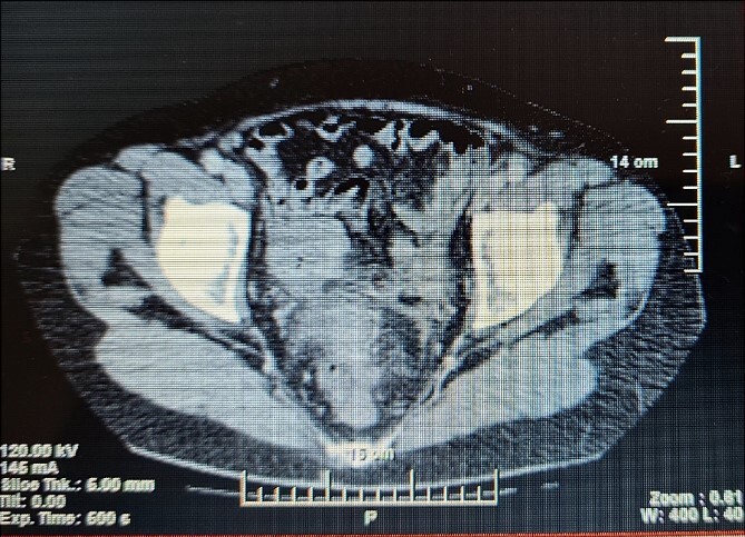 Figure 1
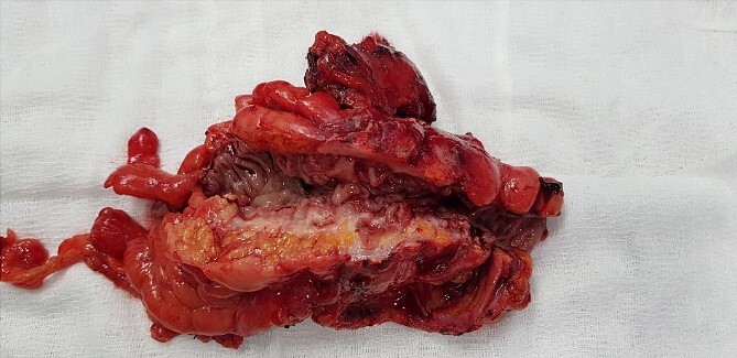 Figure 2
Peer Reviews
No public reviews on file for this paper yet. If you reviewed it on a platform where reviews are public (OpenReview, ICLR, NeurIPS, ICML), you can paste yours below so the community can read it here.
Videos
No videos yet. Explain this paper in a talk, walkthrough, or lecture? Add one.
Taxonomy
TopicsDiverticular Disease and Complications · Gastrointestinal disorders and treatments · Appendicitis Diagnosis and Management
Introduction
Diverticular disease affects a significant percentage of the US population with >00 000 admissions and 1.5 million hospital caries per year, with total annual hospitalizations for acute diverticulitis >7 years increasing by 26% with admission growth of 82% in ages 18 to 44 and 36% between 45 and 74 [1]. Stenosis is a rare complication of acute diverticulitis difficult to distinguish from carcinoma and with some risk of obstruction. The differentiation of benign stenosis from malignant one is extremely difficult and of essential importance given the different therapeutic behavior. A cohort study of 40 496 patients with diverticulitis found a significantly higher incidence of colon cancer in the group with diverticulitis (4.3%) compared to the group without diverticulitis (2.3%)(P < .001) [2]. Therefore these patients should undergo operative treatment despite the negative histological result for malignancy on endoscopic biopsy. We present a case of sigmoid stenosis caused by diverticulosis, mimicking carcinoma.
Case report
A 63-year- old women was admitted to the surgical department with right abdominal pain radiating to the right inguinal region and low back, and the pain had worsened in the last month. Because of these complains a magnetic resonance imaging was performed on an outpatient basis and a thickening of the sigmoid along a length of ~4 cm was found. The patient tells about a benign education, for which the necessary documentation is missing. Colonoscopy was performed which revealed edema and hyperemia of the mucosa ~20 cm from anorectal line to pseudopolypoid dilated lymphatic vessels as in a malignant process. Three biopsies were performed, the result of which proved the presence of moderately inflamed changes and marked lymphostasis with dilated lymphatics. Given the colonoscopy finding and the inability to rule out a malignant process despite a negative biopsy, a contrast- enhanced computed tomography of the abdomen and pelvis was performed. Examination showed a circumferentially and irregularly thickened intestinal bowel wall up to 15 mm ~ 11 cm from the anorectal line along ~6 cm concentrically stenosing lumen, increasing its density characteristic unevently and mainly on the periphery, compacted perirectal fatty tissue, diverticulosis along the sigmoid course, enlarged, regional lymph nodes up to 14/12 mm (Fig. 1). The blood count showed no abnormalities. Given the suspicions of the presence of a malignant process in the sigmoid, the patient underwent surgery and a thickening was found in the sigmoid area and pericolic adipose tissue with infiltration to the right ovary and right ureter. En block resection of the sigmoid along with the right ovary was performed, which was densely attached to the right ureter. On cutting a macroscopic specimen, the cartilaginous density of the tumor along a length of ~6 cm/d and stenosing the lumen of the intestine was impressed, which confirmed our suspicions of malignancy (Fig. 2). Contrary to our expectations, the histological result proved a complicated colonic diverticulitis with exacerbation, abscessation and spread of the inflammatory process with involvement of the pericolic tissues and right ovary, lymph nodes with mixed reactive lymphadenitis. The patient was followed twice with fibrocolonoscopy and with nuclear magnetic resonance postoperatively, which did not show any pathological changes.
CT of the abdomen shows stenosing of the sigmoid colon.
Macroscopic specimen after resection.
Discussion
The hectic lifestyle, the low culture regarding healthy nutrition are the reason why etiological factors such as low fiber content in food [3], obesity [4], smoking [5], the use of steroidal [6], and nonsteroidal [7] anti-inflammatory drugs are associated with the high incidence of diverticulitis in humans.
Very often, diverticulosis does not cause symptoms and is diagnosed when a complication occurs, as in our case. Stenosis due to fibrosis after past diverticulitis is a rare complication, often found in the presence of subileus manifastations and without history and evidence of previous episodes of diverticulitis [8]. One established, the differentiation of benign from malignant stenosis follows, which in many cases is challenging and unclear until the final histological result of the resected stenotic area is obtained, as described by Nishiyama [8] and is in the case presented by us. A retrospective study of 110 patents with complicated diverticular disease by Hussain et al. [9] reported the presence of phlegmon in 31% of cases and stricture/obstruction in 10%. Sigmoid colon is the most common site for complicated diverticulitis in 91% of patients [9].
A meta analysis performed by Meyer et al. [10] including 50 445 patients with acute diverticulitis found an overall incidence of colorectal cancer of 1.9% with a significantly higher risk in patients with complicated diverticulitis compared to patients with uncomplicated diverticulitis. A systematic review of 30 studies involving 29 348 subjects with acute diverticulitis by Kaoo et al. [11] found colon malignancy in 1.67% with a 1.22% risk of malignancy in patients with uncomplicated diverticulitis and 6.14% in complicated. Tehranian et al. [12] found no significant difference in the incidence of colorectal carcinoma in patients with complicated versus uncomplicated diverticulitis (respectively 5.6% and 7.4%), and in 69.2% the diverticulitis was diagnosed at the same site as the tumor. They concluded that colonoscopy is recommended after the diagnosis of diverticulitis, as significantly more colorectal carcinoma was detected after diverticulitis than among subjects undergoing screening colonoscopy.
Computed tomography remains the method of choice for the diagnosis of both acute diverticulitis and emerging complications with a sensitivity of 99% [13]. In the two cases describe by Nishiyama et al. [8] the computed tomography shows the thickening of the bowel wall, and the biopsy excludes the presence of malignancy. In the case describe by us, in addition to thickening of the wall over a length of ~6 sm/d, there are also enlarged lymph nodes, as described by Nishiyama [8] in one of the cases. Enlarged lymph nodes rather make the surgeon to think in the direction of the presence of a malignant disease, and this is a serious prerequisite for resorting to surgical treatment. Differentiating malignant from benign disease in the presence of a diverticulum near the tumor suggests complicated diverticular disease, but operative treatment and definitive histological verification are needed because a negative biopsy does not rule out colon cancer.
Conflict of interest statement
None declared.
Funding
None declared.
The reference list from the paper itself. Each links out to its DOI / PubMed record.
- 1Etzioni DA , Mack TM, Beart RW, Kaiser AM. Diverticulitis in the United States: 1998-2005: changing patterns of disease and treatment. Ann Surg 2009;249:210–7. 10.1097/SLA.0b 013e 3181952888.19212172 · doi ↗ · pubmed ↗
- 2Mortensen LQ , Burcharth J, Andresen K, et al. An 18-year nationwide cohort study on the association between diverticulitis and colon cancer. Ann Surg 2017;265:954–9. 10.1097/SLA.0000000000001794.27192351 · doi ↗ · pubmed ↗
- 3Ma W , Nguyen LH, Song M, et al. Intake of dietary Fiber, fruits, and vegetables and risk of diverticulitis. Am J Gastroenterol 2019;114:1531–8. 10.14309/ajg.0000000000000363.31397679 PMC 6731157 · doi ↗ · pubmed ↗
- 4Ma W , Jovani M, Liu PH, et al. Association between obesity and weight change and risk of diverticulitis in women. Gastroenterology 2018;155:58–66.e 4. 10.1053/j.gastro.2018.03.057.29614301 PMC 6035062 · doi ↗ · pubmed ↗
- 5Aune D , Sen A, Leitzmann MF, et al. Tobacco smoking and the risk of diverticular disease - a systematic review and meta-analysis of prospective studies. Colorectal Dis 2017;19:621–33. 10.1111/codi.13748.28556447 · doi ↗ · pubmed ↗
- 6Humes DJ , Fleming KM, Spiller RC, West J. Concurrent drug use and the risk of perforated colonic diverticular disease: a population-based case-control study. Gut 2011;60:219–24. 10.1136/gut.2010.217281.20940283 · doi ↗ · pubmed ↗
- 7Strate LL , Liu YL, Huang ES, et al. Use of aspirin or nonsteroidal anti-inflammatory drugs increases risk for diverticulitis and diverticular bleeding. Gastroenterology 2011;140:1427–33. 10.1053/j.gastro.2011.02.004.21320500 PMC 3081980 · doi ↗ · pubmed ↗
- 8Nishiyama N , Mori H, Kobara H, et al. Difficulty in differentiating two cases of sigmoid stenosis by diverticulitis from cancer. World J Gastroenterol 2012;18:3623–6. 10.3748/wjg.v 18.i 27.3623.22826630 PMC 3400867 · doi ↗ · pubmed ↗