Vaginal Bleeding in a Peri-Menopausal Woman
Ambika Shivarajpur, Brian Kohen

TL;DR
A 51-year-old perimenopausal woman with vaginal bleeding was diagnosed with a molar pregnancy using point-of-care ultrasound.
Contribution
This case highlights the utility of transvaginal POCUS in the rapid diagnosis of molar pregnancies in perimenopausal women.
Findings
Transvaginal POCUS revealed findings concerning for a molar pregnancy.
The use of POCUS led to prompt diagnosis and treatment initiation.
Vaginal bleeding in perimenopausal women can be effectively evaluated with POCUS.
Abstract
Point of care ultrasound (POCUS) is a useful modality to initially identify a molar pregnancy. In this case, we describe a 51-year-old perimenopausal woman who presented to the emergency department (ED) with vaginal bleeding. A transvaginal POCUS was performed, revealing findings concerning for a molar pregnancy. These findings led to prompt diagnosis and treatment.
Genes, proteins, chemicals, diseases, species, mutations and cell lines named across the full text — each resolved to its canonical identifier and authoritative record.
Click any figure to enlarge with its caption.
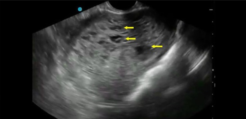 Figure 1
Peer Reviews
No public reviews on file for this paper yet. If you reviewed it on a platform where reviews are public (OpenReview, ICLR, NeurIPS, ICML), you can paste yours below so the community can read it here.
Videos
No videos yet. Explain this paper in a talk, walkthrough, or lecture? Add one.
Taxonomy
TopicsGestational Trophoblastic Disease Studies · Ectopic Pregnancy Diagnosis and Management · Prenatal Screening and Diagnostics
Presentation
A 51-year-old peri-menopausal woman with no significant past medical history presented to the ED with painless vaginal bleeding for 1 day. Her vital signs were within normal limits. On physical examination, the patient had a minimal amount of blood in the vaginal vault. She had no adnexal tenderness to palpation or active bleeding. A transvaginal POCUS revealed an enlarged uterus with cystic, hypoechoic lesions, prompting suspicions of a molar pregnancy (Figure 1, Video S1).
Transvaginal POCUS. Transverse view of an echogenic, enlarged uterus with cystic lesions (arrows) representing the classic“snowstorm” appearance consistent with a complete hydatidiform mole
Evaluation of the adnexa on transvaginal ultrasound was unremarkable. This finding prompted the ED provider to obtain a beta-human chorionic gonadotropin level, which returned greater than 350,000 mIU/mL (< 5 mIU/mL). The patient remained hemodynamically stable and underwent suction curettage by gynecology as well as pathological examination of the uterine contents. The findings showed hydropic villi, and proliferation of cytotrophoblasts and syncytiotrophoblasts. These confirmed a complete molar pregnancy.
Discussion
A hydatidiform mole, also known as a “molar pregnancy”, is an abnormal pregnancy characterized by placental villi with focal swelling, trophoblastic proliferation, and reduplication of genetic material. Hydatidiform moles are distinguished as either complete or partial moles 1. A complete mole is paternally derived, where sperm fertilizes an enucleated ovum 2. Duplication of a haploid sperm results in genotype 46, either XX or XY 2. In contrast, when sperm fertilizes a normal, nucleated ovum, this results in a partial mole with genotype 69, XXY 1, 2. Molar pregnancies occur in approximately 0.5-2 pregnancies per 1,000 2. The most common presenting symptom is vaginal bleeding 3. Risk factors for a molar pregnancy include extremes of ages (<21 or >35 years-old), previous history of molar pregnancy, and nulliparity 2. Generally, molar pregnancies are not viable, and treatment includes molar evacuation 2. The definitive diagnosis of molar pregnancies is made by histological evaluation, which is not immediately available. Given that approximately 3% of hydatidiform moles progress to choriocarcinoma 2, prompt diagnosis and treatment of a molar pregnancy is imperative.
Ultrasound has been used as an initial screening modality for detection of molar pregnancy given its accessibility, accuracy, and rapid identification of key features consistent with the diagnosis 4. Transvaginal ultrasound is more commonly used due to the higher frequency of the endocavitary probe compared to the curvilinear probe used for transabdominal examinations, allowing for a higher resolution image. In both POCUS and comprehensive transvaginal ultrasound, the uterus is viewed in both a transverse and sagittal orientation. It is often requested that the patient empty their bladder prior to scanning as when the bladder is full, it may obscure evaluation of the uterus. Abnormal sonographic findings in molar pregnancy include a focal cystic space within the placenta in patients with partial moles, or an enlarged uterus with multiple hypoechoic cystic lesions in patients with complete molar pregnancy. The classic description of complete molar pregnancy on ultrasound is a “snowstorm” appearance of the uterus due to the numerous hypoechoic cystic lesions present. In this case of complete molar pregnancy, the use of transvaginal POCUS led to prompt diagnosis and treatment.
Disclosures
All authors report no disclosures related to this work.
Patient Consent
The authors obtained informed consent from the patient. The patient gave consent to use de-identified images, videos, and health information for the purpose of publishing in this scientific journal.
Supplementary Material
Video S1Transvaginal POCUS demonstrating an enlarged uterus with cystic lesions consistent with a complete molar pregnancy.
The reference list from the paper itself. Each links out to its DOI / PubMed record.
- 1Berkowitz R S Goldstein D P Clinical practice. Molar pregnancy N Engl J Med 2009360161639168410.1056/NEJ Mcp 0900696.19369669 · doi ↗ · pubmed ↗
- 2Lurain J R Gestational trophoblastic disease I: epidemiology, pathology, clinical presentation and diagnosis of gestational trophoblastic disease, and management of hydatidiform mole Am J Obstet Gynecol 2010203653154010.1016/j.ajog.2010.06.07320728069 · doi ↗ · pubmed ↗
- 3Heaton H A Tintinalli JE Ma OJ Ectopic pregnancy and emergencies in the first 20 weeks of pregnancy Tintinalli’s Emergency Medicine: A Comprehensive Study Guide 9Mc Graw Hill 20209898
- 4Newhouse I Spacey A Scragg B Szczepura K The diagnostic value and accuracy of ultrasound in diagnosing hydatidiform mole: A systematic review and meta-analysis of the literature. Radiography (Lond)202228489790510.1016/j.radi.2022.06.00535785640 · doi ↗ · pubmed ↗