Neuroradiological Correlation of the Lateral Third Periventricular, Pituitary Gland and Stalk, Pineal Gland, Cerebral Aqueduct, and Foramen Magendie and Luschka With Intraventricular Neuroendoscopic Images: A Case Series
Zaitun Zakaria, Muhammad Ihfaz Ismail, Song Yee Ang, Zamzuri Idris

TL;DR
This case series explores how neuroendoscopic images during surgery compare to neuroradiological images of brain structures in pediatric patients.
Contribution
The study provides a direct correlation between intraoperative neuroendoscopic views and radiological images of specific brain regions.
Findings
Intraoperative neuroendoscopic images differ from radiological images, requiring careful navigation.
The study highlights normal and abnormal findings in the ventricular system and surrounding structures.
Anatomical landmarks like the pituitary gland and cerebral aqueduct are crucial for safe neuroendoscopic navigation.
Abstract
Neuroendoscopy procedures in pediatrics have expanded beyond the endoscopic third ventriculostomy. As such, a direct and angled endoscope allows further visualization around the corner, capturing the surrounding anatomy. Intraoperative live images look different than radiological images. Hence, in this single institutional experience, we correlate neuroradiology images with intraoperative intraventricular endoscopic views of the third-fourth ventricle, pituitary, pineal gland, cerebral aqueduct, and foramen magendie and luschka. Our collective case series reveals a few interesting case scenarios of normal and abnormal findings during the procedure. Careful navigation of the neuroendoscope is crucial to prevent injury to the neurovascular bundle. A close relationship with normal anatomy from radiological imaging is necessary to prevent it from getting lost once inside the ventricular…
Genes, proteins, chemicals, diseases, species, mutations and cell lines named across the full text — each resolved to its canonical identifier and authoritative record.
Click any figure to enlarge with its caption.
 Figure 1
 Figure 2
 Figure 3
 Figure 4
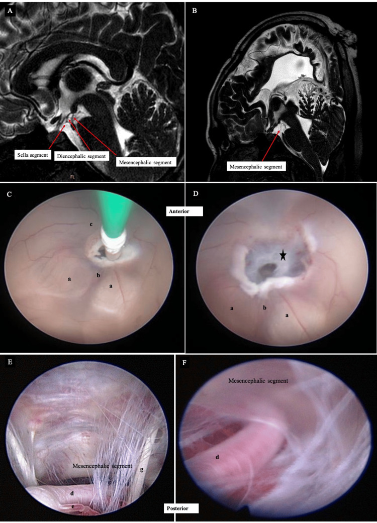 Figure 5
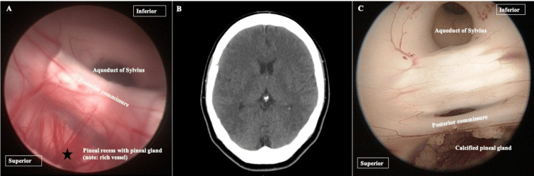 Figure 6
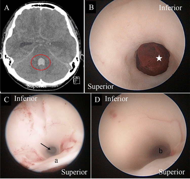 Figure 7
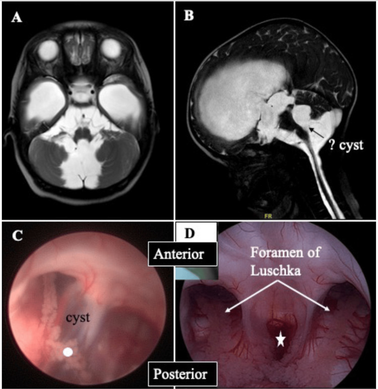 Figure 8
Peer Reviews
No public reviews on file for this paper yet. If you reviewed it on a platform where reviews are public (OpenReview, ICLR, NeurIPS, ICML), you can paste yours below so the community can read it here.
Videos
No videos yet. Explain this paper in a talk, walkthrough, or lecture? Add one.
Taxonomy
TopicsCerebrospinal fluid and hydrocephalus · Fetal and Pediatric Neurological Disorders · Traumatic Brain Injury and Neurovascular Disturbances