Correction: Experience with aortic arch inclusion technique using artificial blood vessel for type A aortic dissection: an application study
Qingfeng Li, Bin Li, Shuqiang Xi, Zhaobin Li, Zhe Zhu, Zeyue Jin, Fan Yang, Lei Liu

Abstract
Genes, proteins, chemicals, diseases, species, mutations and cell lines named across the full text — each resolved to its canonical identifier and authoritative record.
Click any figure to enlarge with its caption.
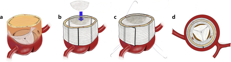 Figure 1
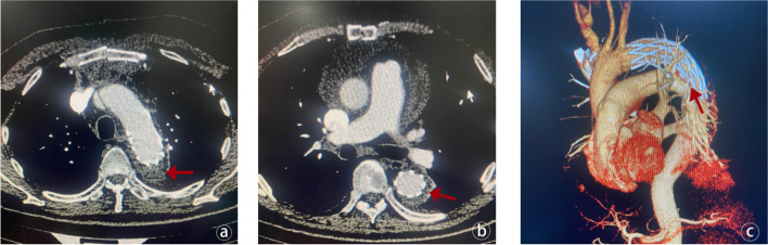 Figure 2
Peer Reviews
No public reviews on file for this paper yet. If you reviewed it on a platform where reviews are public (OpenReview, ICLR, NeurIPS, ICML), you can paste yours below so the community can read it here.
Videos
No videos yet. Explain this paper in a talk, walkthrough, or lecture? Add one.
Taxonomy
TopicsAortic Disease and Treatment Approaches · Aortic aneurysm repair treatments · Cardiac Valve Diseases and Treatments
Correction: J Cardiothorac Surg 19, 189 (2024)
https://doi.org/10.1186/s13019-024-02741-8
Following the publication of the original article [1], Figs 2 and 3 were wrongly published; The figure published as Fig. 3 should actually be Fig. 2. Additionally, the figure originally intended to be presented as Figure 3, which contains vital imaging data pertinent to the study was missing. The figures 2 and 3 should have appeared as shown below.
Fig. 2. Schematic diagram of modified sandwich method of aortic root reinforcement. (a) Semi-perspective diagram of aortic root. (b) The aortic root is sutured and reinforced and a spacer is added. (c) Use the “modified sandwich” method for full-thickness transmural suturing (pay attention to fully exposing the left and right coronal openings). (d) Top view of aortic root after reinforcementFig. 3Assessment of stent and thrombus status after TAAR. (a) The red arrow indicates that the thrombus in the false lumen of the aortic arch has been thrombosed after surgery, and the stent is in good shape. (b) The yellow arrow indicates that the thrombus in the false lumen of the descending aortic stent has been completely thrombosed after surgery, the stent is in good shape, and there is no endoleak. (c) The 3D reconstruction of the patient’s aortic CTA demonstrates the morphology of the stent
The original article has been corrected.
The reference list from the paper itself. Each links out to its DOI / PubMed record.