Lipoma Arising in the Eustachian Tube
Takahiro Inoue, Takumi Kumai, Tomoki Yoshizaki, Miki Takahara

TL;DR
A rare case of a lipoma in the eustachian tube is reported, highlighting its diagnosis and treatment.
Contribution
This paper presents a rare clinical case of a lipoma in the eustachian tube and discusses its diagnosis and surgical removal.
Findings
A 62-year-old female was diagnosed with a lipoma in the eustachian tube using nasal endoscopy and MRI.
The tumor was successfully removed via a transnasal endoscopic approach.
MRI is particularly useful for diagnosing lipomas in the eustachian tube.
Abstract
Lipomas are among the most common soft tissue tumors. Surgical removal of lipoma is considered if the patient has symptoms or cosmetic challenges. Lipomas that develop from any fat tissue in the body and involve the eustachian tube are extremely rare. Herein, we report the case of a patient with a lipoma arising in the eustachian tube. We also summarized the literature on tumors originating from the eustachian tubes. A 62-year-old female presented to our department with a five-year history of left nasal congestion. Nasal endoscopy revealed a tumor in the left eustachian tube. The tumor was considered a lipoma on computed tomography (CT) and magnetic resonance imaging (MRI) and was removed using a transnasal endoscopic approach. Nasal endoscopy and radiologic imaging can be used to detect tumors in the nasopharynx, including the eustachian tubes. Magnetic resonance imaging is…
Genes, proteins, chemicals, diseases, species, mutations and cell lines named across the full text — each resolved to its canonical identifier and authoritative record.
Click any figure to enlarge with its caption.
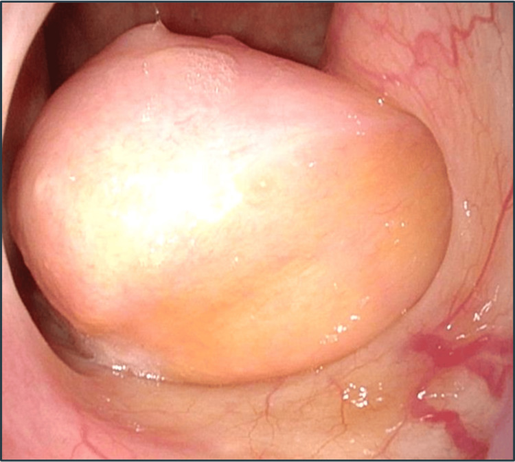 Figure 1
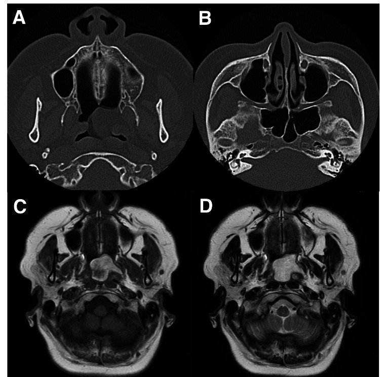 Figure 2
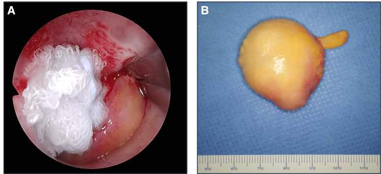 Figure 3
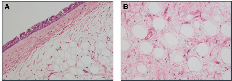 Figure 4
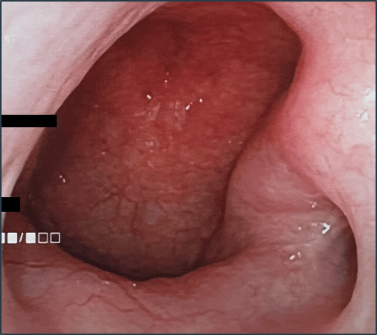 Figure 5
| Tumor | Number |
| Carcinoma | 21 |
| Squamous cell carcinoma/transitional cell carcinoma | 14 |
| Mucoepidermoid carcinoma | 6 |
| Medullary carcinoma | 1 |
| Dermoid cyst | 20 |
| Melanin-pigmented oncocytic metaplasia | 12 |
| Lipoma | 9 |
| Teratoma | 9 |
| Melanoma | 8 |
| Chondroma | 7 |
| Sarcoma | 3 |
| Leiomyoma | 1 |
| Total | 90 |
| Case | Year | Author | Age | Sex | Symptoms | OME | Tumor size (mm) | Follow-up (month) |
| 1 | 2008 |
Park et al. [ | 42 | Female | Aural fullness, hearing loss | + | 10 × 10 | 3 |
| 2 | 2011 |
Liu et al. [ | 34 | Female | Aural fullness, tinnitus, nasal congestion | + | 16 × 24 | 5 |
| 3 | 2012 |
Fuji et al. [ | 29 | Female | Nasal congestion | + | 27 × 10 | N/A |
| 4 | 2013 |
Thakur et al. [ | 50 | Female | Nasal congestion | - | 40 × 40 | 6 |
| 5 | 2016 |
Aydin et al. [ | 43 | Male | Otalgia, hearing loss | + | 4 × 5 | 6 |
| 6 | 2016 |
Dabiri et al. [ | 47 | Male | Aural fullness, otalgia | + | 15 × 8 | 12 |
| 7 | 2022 |
Al Zaabi et al. [ | 27 | Male | Aural fullness, tinnitus, hearing loss | + | 10 × 8 | 36 |
| 8 | 2022 |
Ko et al. [ | 49 | Female | Tinnitus, nasal congestion | + | 7 × 7 | 12 |
| 9 | 2023 | Present case | 62 | Female | Nasal congestion, aural fullness | - | 35 × 30 | 12 |
Peer Reviews
No public reviews on file for this paper yet. If you reviewed it on a platform where reviews are public (OpenReview, ICLR, NeurIPS, ICML), you can paste yours below so the community can read it here.
Videos
No videos yet. Explain this paper in a talk, walkthrough, or lecture? Add one.
Taxonomy
TopicsTumors and Oncological Cases · Teratomas and Epidermoid Cysts · Ear and Head Tumors
Introduction
Lipoma is the most common benign tumor that develops from the mesenchymal connective tissue and is mostly asymptomatic [1]. Only 15% of lipomas are located in the head and neck region [2], mostly in the posterior neck [3]. Moreover, involvement of the eustachian tube in lipoma is extremely rare [4]. Herein, we report the case of a patient with a lipoma arising in the eustachian tube.
Case presentation
A 62-year-old female visited our department with a five-year history of left nasal congestion. Nasal endoscopy revealed a smooth-surfaced tumor that arose from the left eustachian tube to the nasopharynx (Figure 1).
Finding of nasal endoscopyNasal endoscopy revealed a smooth-surfaced tumor arising from the left eustachian tube to the nasopharynx (35 × 30 mm).
The tympanic membrane showed partial myringosclerosis, but no clear middle ear effusion. No abnormal findings were observed in audiography and tympanometry. Computed tomography (CT) revealed a tumor protruding from the left eustachian tube into the nasopharynx, and no soft tissue shadow was observed in the middle ear, suggesting otitis media with effusion (OME). Magnetic resonance imaging (MRI) demonstrated that the tumor had high signal intensity in T1- and T2-weighted images (Figure 2).
Findings of CT and MRIA: Axial plain CT scan revealed a tumor arising from the left eustachian tube with the same density as the surrounding fat. B: Axial plain CT scan revealed no evidence of OME. C: Axial plain T1-weighted MRI scan revealed a tumor with high signal intensity. D: Axial plain T2-weighted MRI scan revealed high signal intensity.CT: computed tomography, MRI: magnetic resonance imaging, OME: otitis media with effusion
Based on these findings, a lipoma was suspected, and surgical removal via a transnasal endoscopic approach under general anesthesia was performed. The tumor was completely embedded in the eustachian tube; however, it had good mobility. We pulled the tumor toward the nasopharynx and resected its base using scissors. The tumor was then removed from the oral cavity. No bleeding was observed after the resection. The patient was discharged without postoperative complications. The tumor was 35 × 30 mm (Figure 3).
Intraoperative findingsA: The tumor was pulled out into the nasopharynx, and the base of the tumor was excised. B: The tumor removed along with the base (35 × 30 mm).
Histopathological examination revealed that the epithelium consisted of a poorly atypical pseudostratified epithelium composed of mature adipocytes with well-defined borders surrounded by fibrous components (Figure 4).
Histopathological findingsA: Hematoxylin and eosin staining revealed that the epithelium consisted of a poorly atypical pseudostratified layer (original magnification, ×100). B: The tumor was composed of mature adipocytes with well-defined borders surrounded by a fibrous component (original magnification, ×200).
There were no malignant findings, and the tumor was diagnosed as a lipoma. One year after surgery, no recurrence of the tumor or nasal obstruction symptoms were observed (Figure 5).
Findings of nasal endoscopy one year after the surgeryNasal endoscopy in the left nasal cavity revealed no recurrence.
Discussion
Tumors arising from the eustachian tube area are extremely rare. The symptoms induced by blocking the eustachian tube include aural fullness, tinnitus, otalgia, hearing loss, and nasal congestion. CT, MRI, nasal endoscopy, audiography, and tympanometry are required for the diagnosis. Although rare, various types of tumors in the eustachian tubes have been reported. We have summarized the reported cases of tumors arising from the eustachian tube (Table 1) [1,4-16].
In a total of 90 cases, squamous cell carcinoma was the most common malignancy with 14 cases, followed by melanoma with eight cases. Among the benign tumors, dermoid cysts were the most common (20 cases), followed by melanin-pigmented oncocytic metaplasia (12 cases), and lipoma and teratoma (each with nine cases).
Lipomas are benign subcutaneous or submucosal tumors that occur between the ages of 50 and 60 years [17]. The origin of the lipoma in the eustachian tube could be the Ostmann fat pad, which is distributed along the eustachian tube. Lipomas are classified into various histological types, including fibro-, spindle cell, pleomorphic, myo-, angio-, chondroid, myxo-, and osteolipomas [8]. Liposarcoma must be considered if the tumor invades surrounding tissues [1]. Our case may be considered a fibrolipoma owing to a large number of fibrous components with mature adipocytes [18]. Imaging studies are useful for diagnosing lipomas. Because the densities of lipomas and fat are comparable on CT, MRI is more useful for diagnosing lipomas. MRI shows high signals in T1- and T2-weighted images, and the signal is suppressed on T1-weighted images with fat-suppressed contrast-enhanced MRI. Surgical removal involves the treatment of lipomas associated with cosmetic problems or subjective symptoms.
To date, nine cases of lipoma arising in the eustachian tube, including our case, have been reported (Table 2) [1,4-6,8,9,14,15].
Among these patients, 67% were female. The median patient age was 43 years (range: 27-62 years). The disease occurred on the right and left sides in six and three cases, respectively. Although not observed in the present case, OME was present in seven out of nine cases. The tumor in our case obstructed the eustachian tube; however, the patient did not develop OME, possibly because of the high mobility of the tumor. The median follow-up period after surgery was nine months (range: 3-36 years). Nasal congestion was the most common symptom, followed by aural fullness, hearing loss, tinnitus, and otalgia. Endoscopic sinus surgery (ESS) is a feasible minimally invasive approach to the eustachian tubes. All reported tumors, including our case, were removed using ESS, and none recurred.
Conclusions
We report the case of a patient with a lipoma in the eustachian tube. Lipomas in the eustachian tube that cause symptoms such as nasal congestion and aural fullness may require surgical removal. The transnasal endoscopic approach is a feasible procedure to remove lipomas involving the eustachian tube.
The reference list from the paper itself. Each links out to its DOI / PubMed record.
- 1A case of lipoma arising in the eustachian tube Ear Nose Throat J Dabiri J Choufani G Delpierre I Hassid S 0795201610.1177/01455613160950010226829697 · doi ↗ · pubmed ↗
- 2Fibrolipoma of buccal mucosa Contemp Clin Dent Khubchandani M Thosar NR Bahadure RN Baliga MS Gaikwad RN 043201210.4103/0976-237X.95119 PMC 335481222629049 · doi ↗ · pubmed ↗
- 3Lipomas of the head and neck: presentation variability and diagnostic work-up J Laryngol Otol El-Monem MH Gaafar AH Magdy EA 475512020061635914710.1017/S 0022215105004597 · doi ↗ · pubmed ↗
- 4The presence of a lipoma in the Eustachian tube: a case report J Med Case Rep Liu Z Wang D Liu Q 436520112189617610.1186/1752-1947-5-436PMC 3177933 · doi ↗ · pubmed ↗
- 5A case of middle ear effusion due to lipoma on the Eustachian tube Korean J Otorhinolaryngol-Head Neck Surg Park BS Park BG Lee HJ Koo SK 9509532008
- 6Fibrolipoma of eustachian tube resected with endoscopy and polypectomy snare Clin Pharm Therap Fujii T Miyake N Takeuchi K 3334222012 https://mol.medicalonline.jp/archive/search?jo=am 7yakrc&ye=2012&vo=22&issue=4
- 7Primary tumors and tumor-like lesions of the eustachian tube: a systematic review of an emerging entity Eur Arch Otorhinolaryngol Muzzi E Cama E Boscolo-Rizzo P Trabalzini F Arslan E 1723173226920122216014410.1007/s 00405-011-1846-x · doi ↗ · pubmed ↗
- 8Fibrolipoma of the eustachian ostium BMJ Case Rep Thakur JS Saluja M Sharma DR Mohindroo NK 2013201310.1136/bcr-2013-008794 PMC 361855623440992 · doi ↗ · pubmed ↗