Integration of diffusion tensor imaging parameters with mesh morphing for in-depth analysis of brain white matter fibre tracts
Maryam Tayebi, Eryn Kwon, Jerome Maller, Josh McGeown, Miriam Scadeng, Miao Qiao, Alan Wang, Poul Nielsen, Justin Fernandez, Samantha Holdsworth, Vickie Shim, Leigh Potter, Leigh Potter, Paul Condron, Davidson Taylor, Daniel Cornfield, Patrick McHugh, Taylor Emsden

TL;DR
This paper introduces a new method combining mesh morphing and PCA to analyze MRI data without averaging, preserving more information for better group comparisons.
Contribution
A novel technique integrating 3D mesh-morphing and PCA for analyzing diffusion tensor MRI data without averaging.
Findings
The new method clearly distinguished rugby players from controls, which traditional averaging could not achieve.
The approach preserves individual variability in diffusion metrics for more accurate group comparisons.
The method enables visualization of diffusion metric statistics across the whole white matter tract volume.
Abstract
Averaging is commonly used for data reduction/aggregation to analyse high-dimensional MRI data, but this often leads to information loss. To address this issue, we developed a novel technique that integrates diffusion tensor metrics along the whole volume of the fibre bundle using a 3D mesh-morphing technique coupled with principal component analysis for delineating case and control groups. Brain diffusion tensor MRI scans of high school rugby union players (n = 30, age 16–18) were acquired on a 3 T MRI before and after the sports season. A non-contact sport athlete cohort with matching demographics (n = 12) was also scanned. The utility of the new method in detecting differences in diffusion tensor metrics of the right corticospinal tract between contact and non-contact sport athletes was explored. The first step was to run automated tractography on each subject’s native space. A…
Genes, proteins, chemicals, diseases, species, mutations and cell lines named across the full text — each resolved to its canonical identifier and authoritative record.
Click any figure to enlarge with its caption.
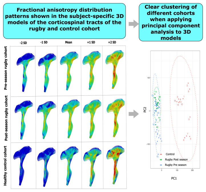 Figure 1
 Figure 2
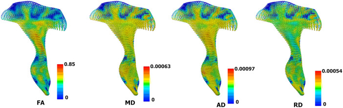 Figure 3
 Figure 4
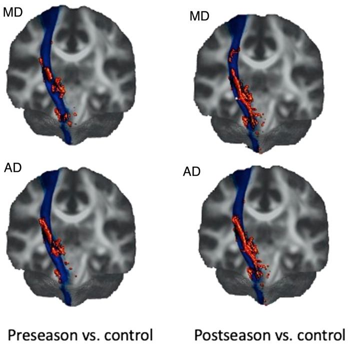 Figure 5
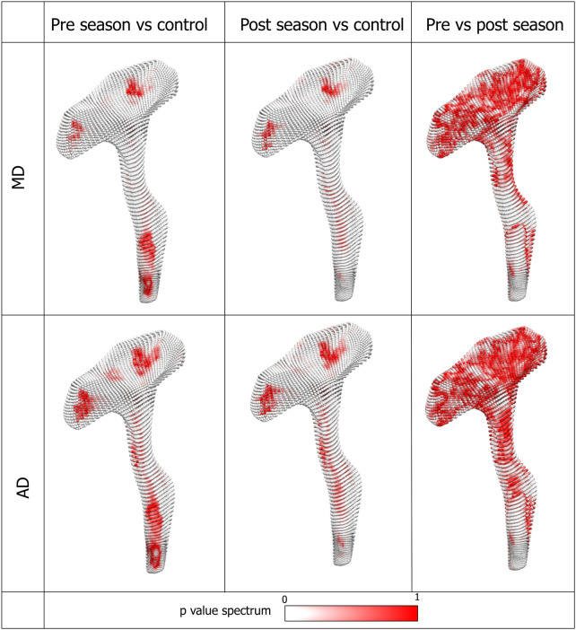 Figure 6
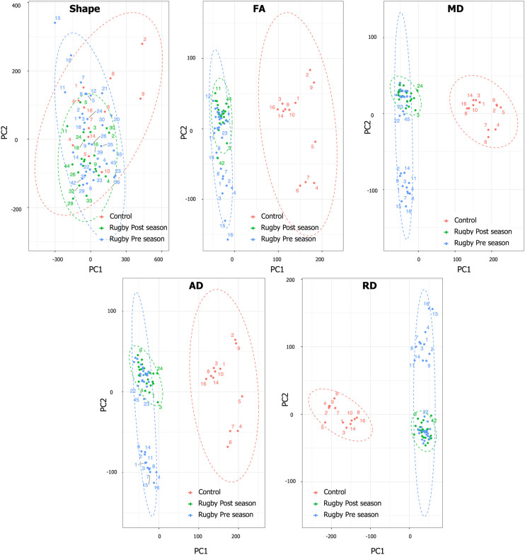 Figure 7
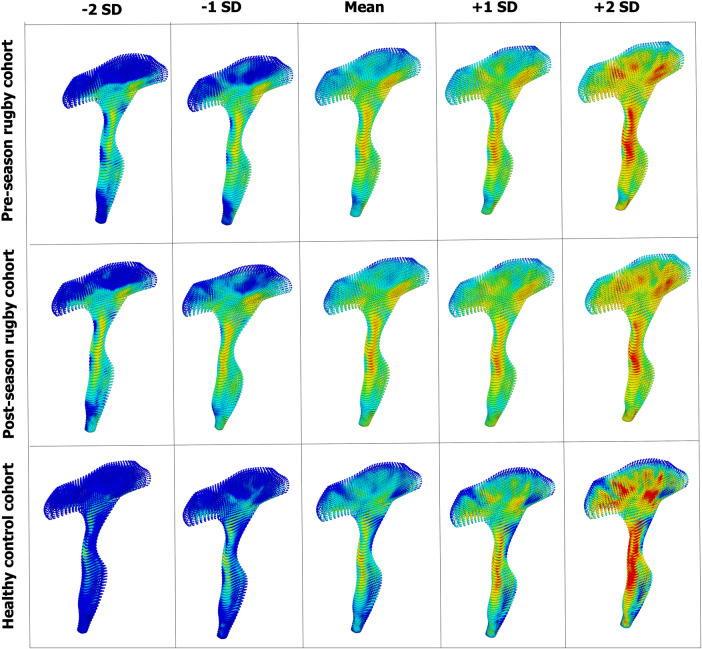 Figure 8
Peer Reviews
No public reviews on file for this paper yet. If you reviewed it on a platform where reviews are public (OpenReview, ICLR, NeurIPS, ICML), you can paste yours below so the community can read it here.
Videos
No videos yet. Explain this paper in a talk, walkthrough, or lecture? Add one.
Taxonomy
TopicsAdvanced Neuroimaging Techniques and Applications · Advanced MRI Techniques and Applications · Fetal and Pediatric Neurological Disorders