Case report: Rapid-onset parkinsonism after a hornet sting
Svetlana Tomic, Milorad Zjalic, Zvonimir Popovic, Zdravka Krivdic Dupan, Marija Heffer, Darija Snajder Mujkic, Dario Mandic, Silva Guljas, Igor N. Petrovic

TL;DR
A rare case of rapid-onset parkinsonism following a hornet sting is reported, highlighting the importance of immunomodulatory treatment to prevent brain damage.
Contribution
This case report adds to the limited literature on neurological syndromes following insect stings and emphasizes the role of immunomodulation.
Findings
A patient developed non-levodopa-responsive parkinsonism and striatal lesions after a hornet sting.
Immunomodulatory treatment led to clinical improvement and reduced brain lesions.
Literature review identified 14 cases, with most showing severe outcomes despite treatment.
Abstract
Neurological manifestations with basal ganglia involvement following Hymenoptera stings are rare and clinically ill-defined conditions. We present a patient with acute parkinsonism non-responsive to levodopa, who developed striatal lesions after a hornet sting. We report his response to immunomodulatory treatment and subsequent clinical and brain magnetic resonance imaging (MRI) follow-up. We also searched the literature for patients with acute extrapyramidal syndromes following an insect sting. Fourteen cases have been published; 12 of them are reviewed here. The majority of cases presented with symmetric akinetic syndrome with axial rigidity and/or gait impairment. Six patients were treated with levodopa and only two of these had a modest response to therapy. Brain MRI/computed tomography scan revealed lesions of the basal ganglia, which resulted in fatal outcome in four patients,…
Genes, proteins, chemicals, diseases, species, mutations and cell lines named across the full text — each resolved to its canonical identifier and authoritative record.
Click any figure to enlarge with its caption.
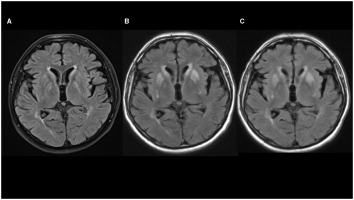 Figure 1
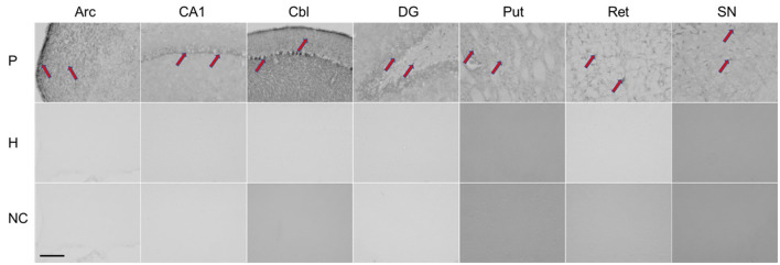 Figure 2
|
|
|
|
|
|
|
|
|
|
|
|
|
|---|---|---|---|---|---|---|---|---|---|---|---|
| Tomic et al. (this case report) | Hornet | 70 | Anaphylaxis 3 days | Freezing of gait; bilateral symmetrical parkinsonism | No signs of lesions | MRI 1—slight bilateral striatum HIS on T1w images, mixed bilateral striatum signal intensity on T2w images with HIS on FLAIR images, without the restriction of diffusion. MRI 3—mixed striatum signal intensity and HIS in ventral putamen on T1w, HIS on T2w and FLAIR images | Normal | Not done | No response to 1,000 mg/day | corticosteroide; PE; rituximab | Partial improvement after the first 2 weeks of treatment with methylprednisolone; after discontinuation of corticosteroides returning of symptoms without further improvement |
| Leopold et al. ( | Wasp | 49 | No anaphylaxis A few hours | Mild resting right hand tremor; bilateral symmetric parkinsonism; postural instability | Symmetrical exaggerated muscle stretch reflexes | MRI 1—bilateral HIS signal in the globus pallidum on T2w images MRI 2—marked destruction of the striatum and pallidum bilaterally | Marked decrease in metabolic activity of the basal ganglia with sparing of the thalamus | Not done | No response to 900 mg/day | PE; IvIg; azathioprine | Stable for 6 months; followed by rapid progression Partial but significant improvement after immunomodulatory therapy |
| Agarwal et al. ( | Honeybee | No data | No data 3 days | Symmetric parkinsonism; short shuffling gait | No signs of lesions | MRI—diffuse HIS on T2w and FLAIR images in bilateral caudate and lentiform nuclei; diffuse effacement of the sulci on the right frontal lobe | No data | Not done | Levodopa/carbidopa | Not given | Symptomatic improvement within 4 weeks; after 3 months parkinsonism had improved |
| Mittal et al. ( | Honeybee | 46 | Anaphylaxis 2 days | Symmetric parkinsonism; hypokinetic dysarthria. | No signs of lesions | No data | No data | Not done | Levodopa/carbidopa | Not given | Symptomatic improvement of speech over the next 2 months; no data for outcome |
| van Agt et al. ( | Wasp | 52 | Anaphylaxis No data | Bradykinesia; rigidity | No signs of lesions | MRI—symmetrical bilateral caudate nuclei and putaminal HIS signal in T1w, T2w and FLAIR images. Iron deposition in the right external capsule in T2w images. | No data | Not done | Levodopa/carbidopa | Not given | Mild improvement with levodopa after 6 months |
| Gallego et al. ( | Wasp | 72 | No anaphylaxis 1 h | Rigidity | Right hemiparesis bilateral Babinski sign; symmetrically brisk tendon jerks | MRI not done; CT scan—low density of both lenticular nuclei, most pronounced in the left globus pallidus | Not done | Bilateral cavitations of the globus pallidus and softening of the putamen and caudate nuclei in subcortical white matter pallor of the myelin | Not given | Not given | Death (72 h after the sting) |
| Castaigne et al. ( | Wasp | No data | No data | No data | No data | Not done | Not done | Severe necrotic lesions in the putamen and less severe lesions in the caudate, thalamus, and red nucleus | Not given | Not given | Coma; death after 55 days |
| Bogolepov et al. ( | Wasp | 51 | Anaphylaxis 1 h | Parkinsonism | No data | Not done | Not done | Bilateral pallidal necrosis with less profound lesions in the substantia nigra | Not given | Not given. | Death after 14 days |
| Laplane et al. ( | Wasp | No data | No anaphylaxis No data | Chorea; buccofacial dyskinesias; compulsive movements | No data | MRI not done; CT scan –bilateral hypodense pallidostriatal lesions | Not done | Not done | Not given | Not given | Gait disorder and myoclonus lasted for several months with slow recovery; compulsive obsessive behavior developed several years later |
| Gale ( | No data | 36 | Anaphylaxis 1 day | Dystonia; symmetric parkinsonism | Brisk tendon reflexes | MRI not done; CT scan—low attenuation in both posterior parietal regions | Not done | Not done | No response to levodopa | Not given | Partial improvement |
| Gale ( | Wasp | 38 | Anaphylaxis A few hours | Akinetic mutism; plastic hypertonicity in the limbs; catatonic posturing | Tendon reflexes symmetrically brisk; Babinski sign bilaterally; clonus | Not done | Not done | Not done | Not given | Not given | Some improvement; regained speech; died 4 months later from pulmonary embolism |
| Sehgal et al. ( | Wasp | No data | No data 2 days | Akinetic-rigid parkinsonism | No data | No data | No data | No data | No data | No data | No data |
| Kumawat et al. ( | Honeybee | 40 | No anaphylaxis 3 days | Symmetric akinetic-rigid parkinsonism; extrapyramidal dysarthria; dystonic posture on all four limbs | No signs of lesions | MRI - in T2w and FLAIR images HIS signals in bilateral basal ganglia and left centrum semioval | Not done | Not done | Not given | Corticosteroide intravenously for 5 days | All symptoms and signs resolved promptly; no data on follow-up |
Peer Reviews
No public reviews on file for this paper yet. If you reviewed it on a platform where reviews are public (OpenReview, ICLR, NeurIPS, ICML), you can paste yours below so the community can read it here.
Videos
No videos yet. Explain this paper in a talk, walkthrough, or lecture? Add one.
Taxonomy
TopicsHistorical Studies in Science · Archaeology and Natural History
Introduction
Neurological manifestations of Hymenoptera stings are rare and mostly present with central or peripheral demyelination syndrome, intracranial hemorrhage and stroke (1–4). In addition, involvement of basal ganglia including pallidostriatal necrosis, coupled with different types of acute-onset parkinsonism, has been previously reported (5–17). Clinical presentation, treatment reaction and disease outcome are still unknown in this rare condition. We present a patient with acute parkinsonism non-responsive to levodopa and striatal lesions after a hornet sting, and document his response to immunomodulatory treatment with clinical and brain MRI follow-up. We also reviewed the literature for similar cases and discuss clinical presentation and prognosis of this rare condition.
Case report
A 70-year-old, right-handed Caucasian male was referred to our department for a second opinion 2 months after he had developed acute neurological symptoms following a hornet sting. On July 22, 2020, the patient had an anaphylactic reaction with hypotension and a brief loss of consciousness soon after a hornet had stung him in the nose. He was followed for the next 24 h in his local general hospital and discharged the next day after complete recovery following antihistamine therapy with desloratadine.
Over the next 3 days, he developed a feeling of malaise and noticed that his walk had become slower and with smaller steps. Desloratadine was discontinued after 8 days, but his gait became even worse with severe hesitation at gait initiation and sudden episodes during walking when he was unable to take a step. In addition, his family members noticed that he was more taciturn and slower to respond to questions. Up until the hornet incident, he had no major health problems except for well-controlled high blood pressure treated with lisinopril.
Diagnostic assessment
Non-contrast axial computed tomography (CT) scan of his brain performed 2 weeks after the hornet sting did not show any signs of acute stroke, tumor, or intracranial hemorrhage. Four days later, non-contrast head MRI showed a slight bilateral striatum hyperintensity signal (HIS) on T1-weighted images, mixed bilateral striatum signal intensity on T2-weighted images with HIS on fluid-attenuated inversion recovery (FLAIR) images, without restriction of diffusion, suggesting petechial blood products corresponding to a T2^*^ susceptibility artifact on SWI sequence (Figure 1A). Levodopa/carbidopa was started and titrated slowly up to 1000 mg per day for the next 3 weeks, but no appreciable improvement was noted.
(A–C) Fluid-attenuated inversion recovery (FLAIR) sequences in axial plane show progression of hyperintensities in basal ganglia [(A) 08/2020; (B) 10/2020; (C) 02/2021].
Two months after the hornet incident, he came to our hospital. On examination, the patient had an impassive face, speech was hypophonic and he manifested palilalia. He had symmetric rigidity of his neck and limbs, and mild symmetric bradykinesia was present in all extremities without tremor. Reflexes were brisk but his plantar responses were normal. On postural reflex testing, he recovered with 2–3 steps. The main problem was his gait with start hesitation and freezing, especially on turning or passing through the door (Supplementary Video 1_Segment 1).
His Movement Disorder Society-Unified Parkinson's Disease Rating Scale (MDS-UPDRS) III score was 35 and he was 2.5. on the Hoehn and Yahr scale. Levodopa/carbidopa treatment was slowly down-titrated and stopped. Lumbar puncture showed a small increase in protein content (0.548 g/L; reference interval 0.170–0.370 g/L) with normal cell numbers. There were no oligoclonal bands or signs of immunoglobulin intrathecal synthesis. Autoimmune antibodies (anti-NMDA-R, anti-AMPA-R1, anti-AMPA-R2, anti-GABAB-R, anti-LGI 1 and anti-CASP-R 2) from cerebrospinal fluid were normal. DaTSPECT showed normal findings. Non-contrast head MRI showed mixed striatum signal intensity on T1-weighted images with hyperintensity in ventral putamen, hyperintensity on T2-weighted and FLAIR images, predominantly in bilateral putamen and caudate nuclei with a patchy restricted diffusion-weighted image signal suggesting necrosis (Figure 1B).
Immunohistochemistry was performed using the patient's serum to evaluate antibodies in brain tissue (for procedure, see additional material). As shown in Figure 2, rat brains reacted with the patient's serum in several regions (nucleus arcuatus, cornu ammonis 1 region of the hippocampus, cerebellum, dentate gyrus of the hippocampus, putamen, reticular formation of the pons and substantia nigra).
Immunohistochemical staining of rat brain slices using human serum as source of the primary antibodies. Red arrows indicate positive staining reaction in different regions of the rat brain. P, patient; H, healthy control; NC, negative controle; Arc, arcuatus; CA1, cornu ammonis 1 region of hippocampus; Cbl, cerebellum; DG, dentate gyrus of hippocampus; Put, putamen; Ret, reticular formation of pons; SN, substantia nigra. Total magnification of individual image 200x, scale 200 micrometers.
With intravenous corticosteroid treatment (methylprednisolone 250 mg/day for 5 days slowly tapered down over the subsequent 2 weeks), marked improvement of gait was observed (Supplementary Video 1_Segment 2). At discharge, the patient still had hesitation while turning, but freezing of gait had ceased, his MDS-UPDRS III score was 18 and he was 2 on the Hoehn and Yahr scale. Continuation of oral pronisone therapy with gradual dose tapering was recommended. Over the next 6 weeks (during which the patient stopped corticosteroid therapy on his own), gradual deterioration of gait with reappearance of freezing occurred. On that time his MDS- UPDRS III score was 30. Five courses of plasma exchange followed by rituximab were administered but without any significant gait improvement.
At that time, head MRI scan (February 28, 2021) showed mixed striatum signal intensity and hyperintensity in the ventral putamen on T1-weighted, hyperintensity on T2-weighted and FLAIR MRI images, suggesting atrophy and gliosis of the striatum, without restriction of diffusion and without contrast enhancement on T1-weighted postcontrast images (Figure 1C). Symptoms remained unchanged and no new symptoms appeared over the next 6 months of follow-up.
Discussion
Basal ganglia necrosis associated with parkinsonism after an insect sting is a rare and ill-defined condition. In our case and the 12 previous cases from the literature (Table 1), presenting features consisted mainly of symmetric akinetic-rigid syndrome which developed within several days of the incident. Resting tremor was rare and mild, and in the majority of cases levodopa response was poor. Instead, the clinical picture consisted of gait impairment including freezing of gait, postural instabilities, and speech disturbances (including palilalia), all of which have been previously recognized as signs of pallidal damage (18). In addition, profound micrographia and akinetic mutism, which were presenting symptoms in one patient each, further support pallidal involvement (19). Interestingly, pyramidal signs including clonus and Babinski sign were noted in five out of eight patients (for the remaining cases, data are lacking). The combination of pyramidal signs coupled with specific, predominantly axial parkinsonism and gait disturbances after an insect sting more closely resemble distinctive, mainly genetically-determined pallido-pyramidal syndrome than Parkinson's disease (20).
MRI was performed in six out of 12 patients, and HIS on T2-weighted and FLAIR images in the region of basal ganglia (striatum, mainly pallidum), were noted in all of them. In three additional cases, CT scans also demonstrated mainly symmetric hypodensity in lenticular nuclei. In our patient, non-contrast brain CT scan and brain MRI performed 2–3 weeks after the hornet sting showed bilateral petechial blood products in the striatum. Non-contrast brain MRI performed 3 months after hornet sting showed signs of bilateral striatal necrosis, and follow-up brain MRI performed after 7 months showed atrophy and gliosis of the striatum. Combining those data with normal DaTSPECT in our case suggest a postsynaptic cause of parkinsonism predominantly due to striatal damage with sparing of the nigrostriatal dopamine bundle.
The etiology of basal ganglia damage in this rare condition is unknown. Hypoxic-ischaemic mechanism due to an exaggerated anaphylaxis, associated hypotension, and eventual prolonged cerebral hypoxia is one possible mechanism. This sequence of events might explain cases with severe hypotension, prolonged loss of consciousness and respiratory failure. Nevertheless, in case presented by Gallego et al. patient presented with stupor 1 h after incident, soon progressed to coma but without significant cardiovascular, electrolytic or respiratory failure (8). In addition, authors stated that histological pattern on necropsy did not suggest a hypoxic-ischaemic nature of basal ganglia damage. In case we presented, as well as in other cases collected here the anaphylactic reaction led to hypotension without or only with short-term loss of consciousness that did not require cardiopulmonary resuscitation, and there was no need for respiratory support suggesting the hypoxic-ischemic mechanism of brain damage unlikely (6, 17).
Given the acute onset of parkinsonism and neuroimaging findings, an immune-mediated early hypersensitivity reaction is most likely. In support of this are evidence for autoimmunity obtained from immunohistochemistry and the excellent treatment response observed in one patient who received corticosteroids in the acute phase, as well as the beneficial but limited clinical response to corticosteroid treatment observed in our case and those reported by Leopold et al. (6). In the latter two cases, immunomodulatory treatment was applied with significant delay. Although we found that antibody reacted with different brain regions (nucleus arcuatus, cornu ammonis 1 region of hippocampus, cerebellum, dentate gyrus of hippocampus, putamen, reticular formation of pons and substantia nigra), only the striatum was damaged by necrosis. Susceptibility of the basal ganglia to toxic and hypoxic lesions has already been described in human and animal studies, and mitochondrial failure with energy deprivation is considered as the main underlying mechanism (21).
Patient perspective
In conclusion, clinicians should be aware of this rare but devastating cause of acute-onset parkinsonism, it‘s specific clinical presentation and clinical course. Although limited, current knowledge suggest that prompt and prolonged immunomodulatory therapy could be a rational therapeutic choice in attempt to prevent irreversible basal ganglia damage.
Data availability statement
The raw data supporting the conclusions of this article will be made available by the authors, without undue reservation.
Ethics statement
Ethical review and approval was not required for the study on human participants in accordance with the local legislation and institutional requirements. Written informed consent from the patients/participants or patients/participants' legal guardian/next of kin was not required to participate in this study in accordance with the national legislation and the institutional requirements. Written informed consent was obtained from the individual(s) for the publication of any potentially identifiable images or data included in this article.
Author contributions
ST: Conceptualization, Funding acquisition, Investigation, Writing—original draft, Writing—review & editing. MZ: Investigation, Writing—original draft, Writing—review & editing. ZP: Investigation, Writing—original draft, Writing—review & editing. ZK: Data curation, Investigation, Writing—original draft, Writing—review & editing. MH: Data curation, Writing—original draft, Writing—review & editing. DS: Data curation, Investigation, Writing—original draft, Writing—review & editing. DM: Data curation, Investigation, Writing—original draft, Writing—review & editing. SG: Data curation, Investigation, Writing—original draft, Writing—review & editing. IP: Conceptualization, Formal analysis, Methodology, Project administration, Resources, Validation, Writing—original draft, Writing—review & editing.
The reference list from the paper itself. Each links out to its DOI / PubMed record.
- 1Riggs JE Ketonen LM Bodensteiner JB Benesch CG. Wasp sting-associated cerebral infarction: a role for cerebrovascular sympathetic innervation. Clin Neuropharmacol. (1993) 16:362–5. 10.1097/00002826-199308000-000098374916 · doi ↗ · pubmed ↗
- 2Romano JT Riggs JE Bodensteiner JB Gutmann L. Wasp sting associated occlusion of the supraclinoid internal carotid artery: implications regarding the pathogenesis of moyamoya syndrome. Arch Neurol. (1989) 46:607–8. 10.1001/archneur.1989.005204200250182730372 · doi ↗ · pubmed ↗
- 3Schoene WC Carpenter SS Behan PO Geschwind N. ‘Onion bulb' formation in the central and peripheral nervous system in association with multiple sclerosis and hypertrophic polyneuropathy. Brain. (1977) 100:755–73. 10.1093/brain/100.4.755204396 · doi ↗ · pubmed ↗
- 4Keller M. Optic neuritis after wasp sting. Klin Monatsbl Augenheilkd. (1995) 206:367–8. 10.1055/s-2008-10354647609388 · doi ↗ · pubmed ↗
- 5Mittal R Munjal S Khurana D Gupta A. Parkinsonism following bee sting: a case report. Case Rep Neurol Med. (2012) 47, 6523. 10.1155/2012/47652323091749 PMC 3474225 · doi ↗ · pubmed ↗
- 6Leopold NA Bara-Jimenez W Hallett M. Parkinsonism after a wasp sting. Mov Disord. (1999) 14:122–7.9918354 10.1002/1531-8257(199901)14:1<122::aid-mds 1020>3.0.co;2-s · doi ↗ · pubmed ↗
- 7van Agt TFA Oliveira KLS Bezerra MER Pedroso JL Franco CMR Melo ES. Acute parkinsonism and basal ganglia lesions after wasp sting. Arq Neuropsiquiatr. (2022) 80:869–70. 10.1055/s-0042-175521536252597 PMC 9703893 · doi ↗ · pubmed ↗
- 8Gállego J Tuñón T Soriano G Delgado G Lacruz F Villanueva JA. Bilateral pallidostriatal necrosis caused by a wasp sting: a clinical and pathological study. J Neurol Neurosurg Psychiatry. (1995) 58:474–6. 10.1136/jnnp.58.4.4747738559 PMC 1073438 · doi ↗ · pubmed ↗