Computer based method for identification of fibrotic scars from electrograms and local activation times on the epi- and endocardial surfaces of the ventricles
Arstanbek Okenov, Timur Nezlobinsky, Katja Zeppenfeld, Nele Vandersickel, Alexander V. Panfilov

TL;DR
This paper introduces a computational method to identify fibrotic scars in the heart using electrical signals from the ventricles, improving detection accuracy in challenging regions.
Contribution
A novel computational method for 3D fibrosis reconstruction using electrogram data from both ventricular surfaces.
Findings
The method successfully detected fibrosis in 204 left ventricle segments with an average error of 0.0±4.3%.
It effectively identified fibrotic scars in the mid-myocardial region, where traditional amplitude-based methods struggle.
Abstract
Cardiac fibrosis stands as one of the most critical conditions leading to lethal cardiac arrhythmias. Identifying the precise location of cardiac fibrosis is crucial for planning clinical interventions in patients with various forms of ventricular and atrial arrhythmias. As fibrosis impedes and alters the path of electrical waves, detecting fibrosis in the heart can be achieved through analyzing electrical signals recorded from its surface. In current clinical practices, it has become feasible to record electrical activity from both the endocardial and epicardial surfaces of the heart. This paper presents a computational method for reconstructing 3D fibrosis using unipolar electrograms obtained from both surfaces of the ventricles. The proposed method calculates the percentage of fibrosis in various ventricular segments by analyzing the local activation times and peak-to-peak amplitudes…
Genes, proteins, chemicals, diseases, species, mutations and cell lines named across the full text — each resolved to its canonical identifier and authoritative record.
Click any figure to enlarge with its caption.
 Figure 1
 Figure 2
 Figure 3
 Figure 4
 Figure 5
 Figure 6
 Figure 7
 Figure 8
 Figure 9
 Figure 10
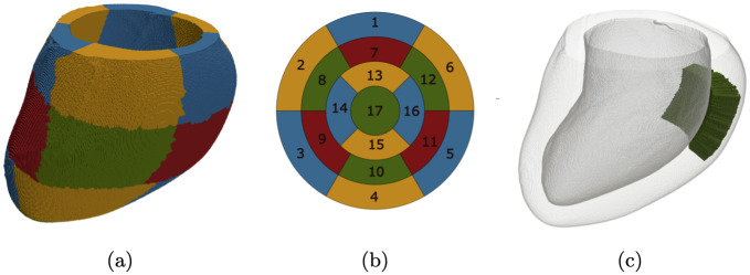 Figure 11
 Figure 12
 Figure 13
 Figure 14
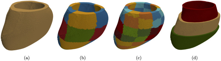 Figure 15
 Figure 16
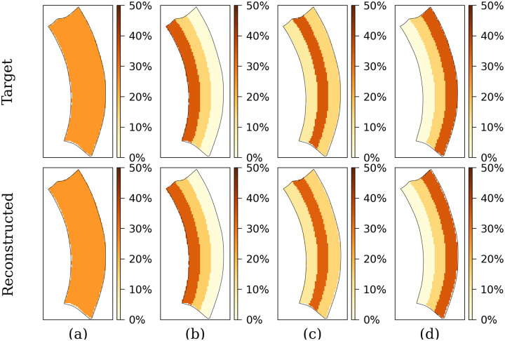 Figure 17
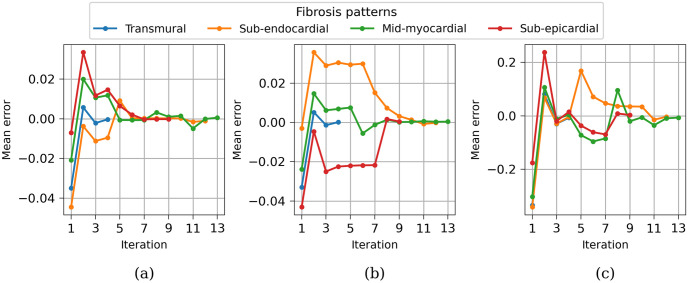 Figure 18
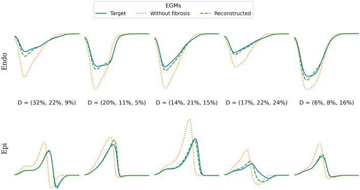 Figure 19
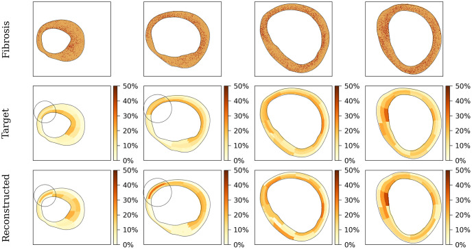 Figure 20
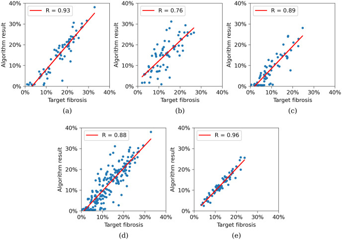 Figure 21
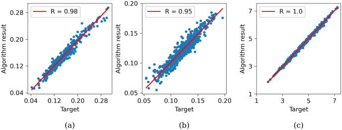 Figure 22
 Figure 23
 Figure 24
 Figure 25
 Figure 26
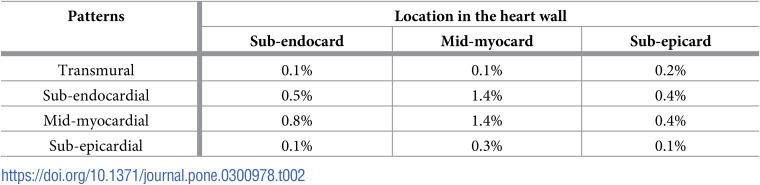 Figure 27
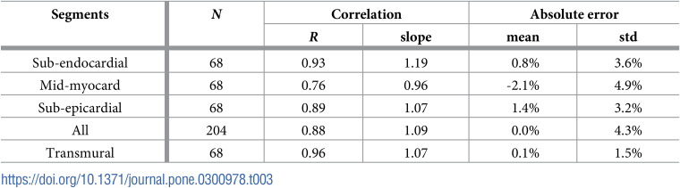 Figure 28
Peer Reviews
No public reviews on file for this paper yet. If you reviewed it on a platform where reviews are public (OpenReview, ICLR, NeurIPS, ICML), you can paste yours below so the community can read it here.
Videos
No videos yet. Explain this paper in a talk, walkthrough, or lecture? Add one.
Taxonomy
TopicsCardiac electrophysiology and arrhythmias · Cardiac Arrhythmias and Treatments · ECG Monitoring and Analysis