Papillary meningioma with prominent flow voids
Mousam Panigrahi, Narendra K. Bodhey, Vandita Y. Singh, Lokesh Nehete

TL;DR
This paper reports a rare case of papillary meningioma with unique MRI features that helped improve surgical outcomes.
Contribution
The novelty lies in highlighting the rare presentation of papillary meningioma with prominent flow voids and increased perfusion on MRI.
Findings
Prominent flow voids and increased perfusion on MRI were observed in a case of papillary meningioma.
These MRI features aided in minimizing intra-operative blood loss and achieving a favorable outcome.
The case highlights the importance of recognizing atypical features for better management and prognosis.
Abstract
This report presents an extremely rare case of papillary meningioma with prominent flow voids and increased perfusion parameters on MRI in a 28-year-old male presenting with headache. This knowledge helped the neurosurgeon to minimise intra-operative blood loss and achieve a favourable post-surgical outcome. A rare case of papillary meningioma and its differentiating features from typical meningiomas have been discussed considering its implications for management as well as prognostication to reduce morbidity and mortality.
Genes, proteins, chemicals, diseases, species, mutations and cell lines named across the full text — each resolved to its canonical identifier and authoritative record.
Click any figure to enlarge with its caption.
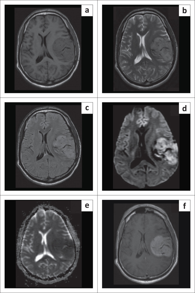 Figure 1
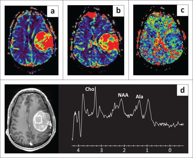 Figure 2
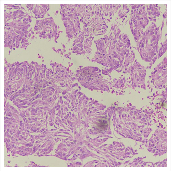 Figure 3
Peer Reviews
No public reviews on file for this paper yet. If you reviewed it on a platform where reviews are public (OpenReview, ICLR, NeurIPS, ICML), you can paste yours below so the community can read it here.
Videos
No videos yet. Explain this paper in a talk, walkthrough, or lecture? Add one.
Taxonomy
TopicsMeningioma and schwannoma management · Head and Neck Surgical Oncology · Neurofibromatosis and Schwannoma Cases