Submicronic-Scale Mechanochemical Characterization of Oxygen-Enriched Materials
Marie Garnier, Eric Lesniewska, Virgil Optasanu, Bruno Guelorget, Pascal Berger, Luc Lavisse, Manuel François, Irma Custovic, Nicolas Pocholle, Eric Bourillot

TL;DR
A new technique called scanning microwave microscopy is shown to effectively measure oxygen concentration and mechanical properties at a very small scale in metal materials.
Contribution
The novel contribution is the calibration and demonstration of scanning microwave microscopy for quantifying oxygen and mechanical properties in metallic materials.
Findings
Scanning microwave microscopy was calibrated using nuclear reaction analysis and nanoindentation.
The technique reliably characterized oxygen-enriched layers on Ti-6Al-4V alloy.
This approach enables indirect quantification of light element diffusion in metals.
Abstract
Conventional techniques that measure the concentration of light elements in metallic materials lack high-resolution performance due to their intrinsic limitation of sensitivity. In that context, scanning microwave microscopy has the potential to significantly enhance the quantification of element distribution due to its ability to perform a tomographic investigation of the sample. Scanning microwave microscopy associates the local electromagnetic measurement and the nanoscale resolution of an atomic force microscope. This technique allows the simultaneous characterization of oxygen concentration as well as local mechanical properties by microwave phase shift and amplitude signal, respectively. The technique was calibrated by comparison with nuclear reaction analysis and nanoindentation measurement. We demonstrated the reliability of the scanning microwave technique by studying thin…
Genes, proteins, chemicals, diseases, species, mutations and cell lines named across the full text — each resolved to its canonical identifier and authoritative record.
Click any figure to enlarge with its caption.
 Figure 1
 Figure 2
 Figure 3
 Figure 4
 Figure 5
 Figure 6
 Figure 7
 Figure 8
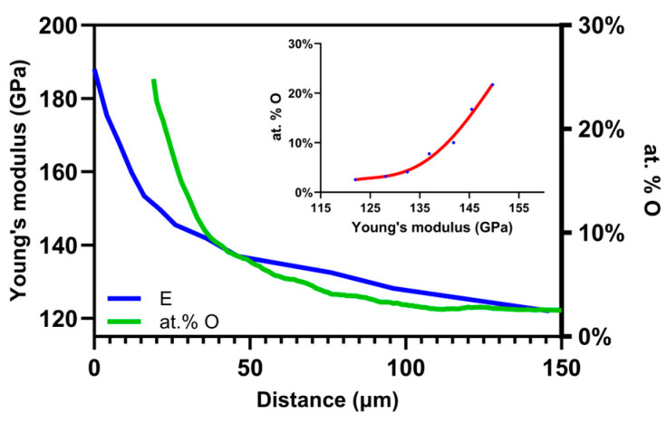 Figure 9
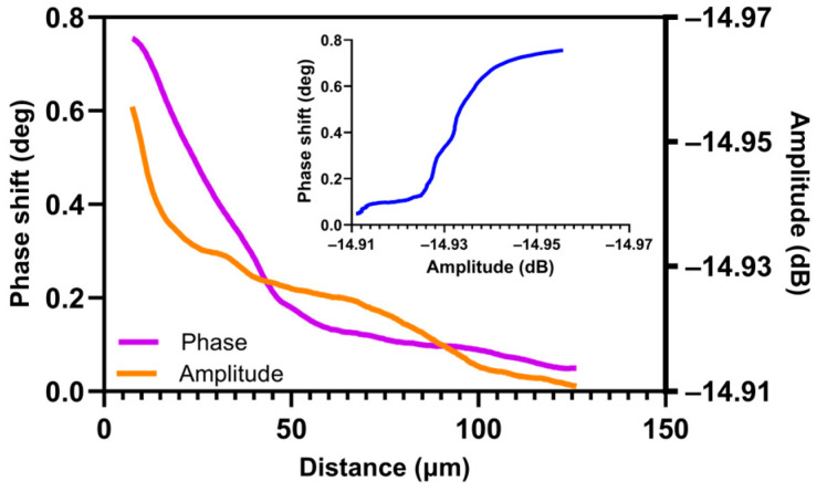 Figure 10
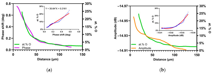 Figure 11
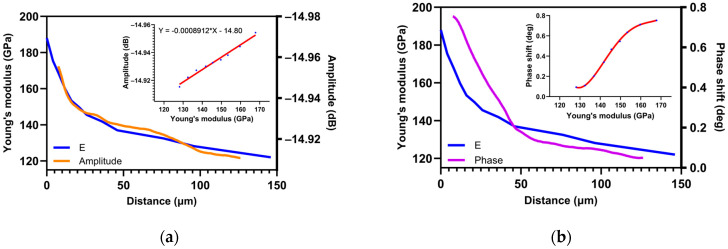 Figure 12
Peer Reviews
No public reviews on file for this paper yet. If you reviewed it on a platform where reviews are public (OpenReview, ICLR, NeurIPS, ICML), you can paste yours below so the community can read it here.
Videos
No videos yet. Explain this paper in a talk, walkthrough, or lecture? Add one.
Taxonomy
TopicsForce Microscopy Techniques and Applications · Near-Field Optical Microscopy · Ultrasonics and Acoustic Wave Propagation