Arterial Calcification Disappearance in Breast Imaging: A Key Indicator for Transition to Invasive Ductal Carcinoma
Arisa Sato, Tomoyuki Fujioka, Iichiroh Onishi, Emi Yamaga, Leona Katsuta, Kazunori Kubota, Yuichi Kumaki, Toshiyuki Ishiba, Goshi Oda, Ukihide Tateishi

TL;DR
A case study shows that the disappearance of arterial calcification in mammograms can signal the transition from a benign breast mass to invasive ductal carcinoma.
Contribution
This case highlights arterial calcification changes as a novel diagnostic indicator for breast cancer progression.
Findings
Gradual disappearance of arterial calcification around a breast mass was observed over time.
The absence of arterial calcification around tumor-associated arteries was noted in invasive ductal carcinoma.
Monitoring calcification changes may improve breast cancer diagnosis beyond traditional imaging methods.
Abstract
A woman in her 70s, initially suspected of having fibroadenoma due to a well-defined mass in her breast, underwent regular mammography and ultrasound screenings. Over several years, no appreciable alterations in the mass were observed, maintaining the fibroadenoma diagnosis. However, in the fourth year, an ultrasound indicated slight enlargement and peripheral irregularities in the mass, even though the mammography images at that time showed no alterations. Interestingly, mammography images over time showed the gradual disappearance of previously observed arterial calcification around the mass. Pathological examination eventually identified the mass as invasive ductal carcinoma. Although the patient had breast tissue arterial calcification typical of atherosclerosis, none was present around the tumor-associated arteries. This case highlights the importance of monitoring arterial…
Genes, proteins, chemicals, diseases, species, mutations and cell lines named across the full text — each resolved to its canonical identifier and authoritative record.
Click any figure to enlarge with its caption.
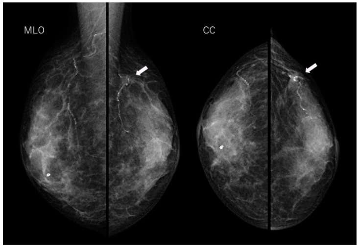 Figure 1
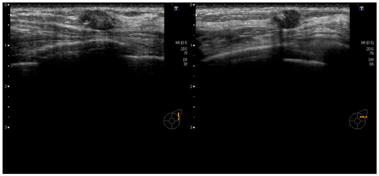 Figure 2
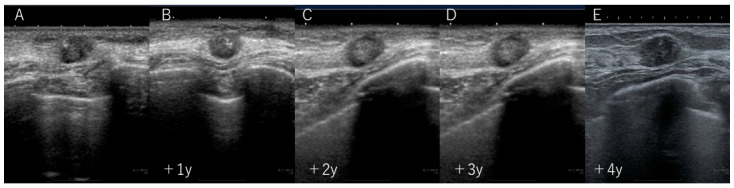 Figure 3
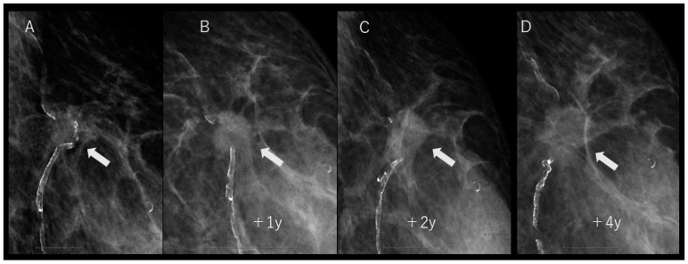 Figure 4
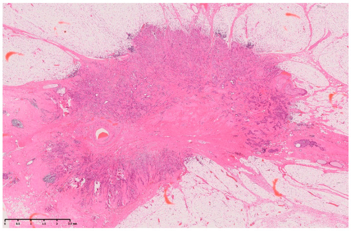 Figure 5
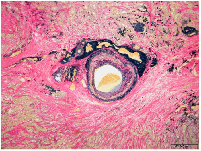 Figure 6
Peer Reviews
No public reviews on file for this paper yet. If you reviewed it on a platform where reviews are public (OpenReview, ICLR, NeurIPS, ICML), you can paste yours below so the community can read it here.
Videos
No videos yet. Explain this paper in a talk, walkthrough, or lecture? Add one.
Taxonomy
TopicsJudicial and Constitutional Studies · Political Philosophy and Ethics · American Constitutional Law and Politics
Breast cancer is one of the most common forms cancers in women, and early detection plays a crucial role in its treatment and prognosis. Common screening methods include ultrasound and mammography, which are widely used to detect abnormalities in breast tissue [1]. Despite the effectiveness of these methods, some cases progress in unusual ways. In our study, we report a rare instance observed during follow-up, in which arterial calcification, initially identified in mammography, disappeared. This report not only shares this unique case, but also explores the potential mechanisms underlying this phenomenon through an extensive literature review. A key aspect of our study includes a detailed comparison between breast imaging and pathological findings, shedding light on the correlations and discrepancies between these diagnostic modalities.
The patient was a 70-year-old woman who had previously been treated for hepatitis C, but was not undergoing any treatment at the time, including medication. She had no other significant medical history, and there was no family history of breast or ovarian cancer. She underwent regular screenings, including ultrasound and mammography. Initial examinations revealed a well-defined, oval-shaped mass with clear borders in her breast, which raised suspicion of fibroadenoma (Figure 1 and Figure 2). Over several years, annual ultrasound and mammography screenings showed no appreciable alterations in the size or shape of the mass, sustaining the fibroadenoma diagnosis.
During the fourth-year follow-up, although there was no apparent change in its size, a subsequent ultrasound examination revealed slight enlargement and irregularities at the periphery of the mass (Figure 3). Surprisingly, a review of previous mammography images revealed the disappearance of pre-existing arterial calcifications within and around the mass. Furthermore, upon examining the mammography images over time, a gradual disappearance of calcifications was observed (Figure 4).
Pathological examination results confirmed that the breast mass was an invasive ductal carcinoma (Figure 5). Surgical pathology also indicated thick blood vessels within the tumor, suggesting a correlation with the initially observed calcified artery. However, there were no clear signs of tumor invasion into the blood vessels. Notably, although the patient had calcifications (atherosclerosis) in the arteries of her normal breast tissue, calcifications were not observed within or around the tumor-associated arteries (Figure 6).
This case, although initially diagnosed as fibroadenoma and observed over time, eventually presented with invasive ductal carcinoma. Although the alteration in the mass’s size over time was limited, the disappearance of arterial calcification within and around the tumor was observed. Although rare, there have been past reports of cases where calcification within the ducts disappeared with the emergence of invasive cancer [2,3], so in this case, the disappearance of calcification could be related to the progression of breast cancer, and the potential mechanisms of this were examined. We considered the mechanism of tumor invasion into the arterial wall as a cause of calcification disappearance. However, pathological evidence to support this hypothesis was not found.
As the malignancy of a tumor increases, blood perfusion within the tumor becomes heterogeneous. This is due to the presence of regions with high blood perfusion, moderate perfusion, low perfusion, and zero perfusion or necrotic tumor zones [4]. Such changes in blood flow are attributed to alterations in vascular structure caused by rapid tumor growth. Normally, arterial wall calcification occurs in a stable blood flow condition. However, as breast cancer progresses, an increase in blood perfusion heterogeneity may lead to the dissolution of existing calcifications. Additionally, the tumor may secrete cytokines that promote angiogenesis, which could also influence the disappearance of calcification [5].
Although not breast cancer, a previous case reported calcification loss in patients with glioma, owing to local malignant alterations. The authors hypothesized that, in the presence of malignant tumors, calcifications may disappear due to a decrease in microenvironmental pH [6]. This factor could have been involved in the arterial wall calcification observed in our patient. It is important to note, however, that our analysis of the mechanisms behind this phenomenon is purely speculative and based on a literature review. Proving such mechanisms conclusively is challenging, and there are inherent limitations in confirming these hypotheses without further detailed research and case accumulation.
Calcification within the breast is a common finding, and mammography is the most effective method for its detection. Additionally, although calcification within the mammary ducts can occur in benign and malignant tumors, their nature can often be inferred from their distribution and morphology [7]. Calcification can also arise from fibroadenoma in the interstitial tissues and from arteriosclerotic changes in the arterial walls. A recent study indicated that mammography can be used to detect arterial wall calcification, which may have potential implications in the risk assessment of cardiovascular events [8]. Calcification detected by mammography can arise from various causes; therefore, interpreting the cause of calcification becomes a crucial factor for diagnosis.
When monitoring cases using mammography, the primary focus is often on changes in the size and shape of the tumor and the appearance and rate of calcification. However, this report emphasizes the clinical significance of observing reductions in arterial calcification within and around tumors. Changes in arterial calcifications may serve as a crucial indicator for the early detection of breast cancer. It is essential to be aware of this possibility in clinical practice and further investigate this phenomenon in future research.
The reference list from the paper itself. Each links out to its DOI / PubMed record.
- 1Ohnuki K. Tohno E. Tsunoda H. Uematsu T. Nakajima Y. Overall assessment system of combined mammography and ultrasound for breast cancer screening in Japan Breast Cancer 20212825426210.1007/s 12282-020-01203-y 33389614 PMC 7925504 · doi ↗ · pubmed ↗
- 2Paolini B. Leddy R. Irshad A. Disappearing grouped breast calcifications: An ominous sign Radiol. Case Rep.2020152453245810.1016/j.radcr.2020.08.06233005283 PMC 7519265 · doi ↗ · pubmed ↗
- 3Seymour H.R. Cooke J. Given-Wilson R.M. The significance of spontaneous resolution of breast calcification Br. J. Radiol.1999723810.1259/bjr.72.853.1034168210341682 · doi ↗ · pubmed ↗
- 4Singh M. Modified Pennes bioheat equation with heterogeneous blood perfusion: A newer perspective Int. J. Heat Mass Transf.202421812469810.1016/j.ijheatmasstransfer.2023.124698 · doi ↗
- 5Esquivel-Velázquez M. Ostoa-Saloma P. Palacios-Arreola M.I. Nava-Castro K.E. Castro J.I. Morales-Montor J. The role of cytokines in breast cancer development and progression J. Interferon Cytokine Res.20153511610.1089/jir.2014.002625068787 PMC 4291218 · doi ↗ · pubmed ↗
- 6Halpin S. Kingsley D. Disappearance of cerebral calcification as a sign of tumor growth AJNR Am. J. Neuroradiol.1993141191228427072 PMC 8334468 · pubmed ↗
- 7Nyante S.J. Lee S.S. Benefield T.S. Hoots T.N. Henderson L.M. The association between mammographic calcifications and breast cancer prognostic factors in a population-based registry cohort Cancer 201712321922710.1002/cncr.3028127683209 PMC 5287030 · doi ↗ · pubmed ↗
- 8Osman M. Regner S. Osman K. Shahan C. Kheiri B. Kadiyala M. Sokos G. Sengupta P.P. Shapiro M.D. Michos E.D. Association between breast arterial calcification on mammography and coronary artery disease: A systematic review and meta-analysis J. Women’s Health 2022311719172610.1089/jwh.2020.873333826862 PMC 9836700 · doi ↗ · pubmed ↗