Genome sequence of Soos: a siphovirus of the CP cluster infecting Gordonia rubripertincta
Reese M. Adams, Holly A. Britton, Emily D. Bruce, Yucita De La Paz, Emily N. Kratz, Emma J. Pfeifer, Daisy E. Priddy, Brooklyn I. Schotter, Wyatt A. Stuffle, Jordyn Wagner, Meredith R. Weiss, Danielle K. Watt, Pamela L. Connerly, Elizabeth E. Rueschhoff

TL;DR
This paper describes the genome of a new siphovirus called Soos, isolated from soil and infecting a specific bacterium.
Contribution
The paper presents the genome sequence of Soos, a rare member of the CP cluster of actinobacteriophages.
Findings
Soos has a 57,509 bp circularly permuted genome.
The genome encodes 87 predicted protein-coding genes.
Soos is one of only three known phages in cluster CP.
Abstract
Novel actinobacteriophage Soos was isolated and purified from Southern Indiana soil using host Gordonia rubripertincta NRRL B-16540. Sequencing revealed a 57,509 bp circularly permuted genome encoding 87 predicted protein-coding genes. Soos is only the third phage in cluster CP, along with phages Clawz and Sting.
Genes, proteins, chemicals, diseases, species, mutations and cell lines named across the full text — each resolved to its canonical identifier and authoritative record.
Click any figure to enlarge with its caption.
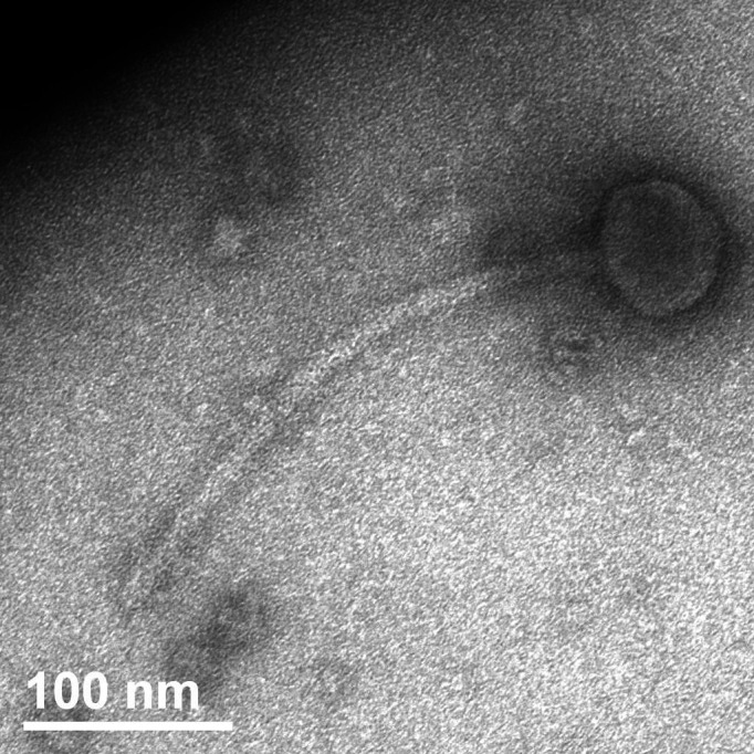 Fig 1
Fig 1- —Indiana University Southeast (IU Southeast)
Peer Reviews
No public reviews on file for this paper yet. If you reviewed it on a platform where reviews are public (OpenReview, ICLR, NeurIPS, ICML), you can paste yours below so the community can read it here.
Videos
No videos yet. Explain this paper in a talk, walkthrough, or lecture? Add one.
Taxonomy
TopicsBacteriophages and microbial interactions · Microbial infections and disease research · Plant and Fungal Interactions Research
ANNOUNCEMENT
Gordonia species are commonly found in soil and water and have occasionally caused illness (1). To date, over 720 Gordonia phages have been sequenced (2).
Soos, a novel bacteriophage that infects Gordonia rubripertincta NRRL B-16540*,* was isolated from a soil sample from Jeffersonville, Indiana, USA (Global Positioning System [GPS] coordinates 38.34446 N, 85.81798 W) using standard protocols (3, 4). The sample was washed in peptone yeast calcium agar (PYCa) media, which was then filtered through a 0.22 µm filter. G. rubripertincta was added to the filtrate and shaken at 250 rpm for 5 days at 26°C. An aliquot of enriched sample was filtered (0.22 µm), mixed with G. rubripertincta, and plated by soft-agar overlay. Incubation at 26°C for 2 days revealed 1 mm diameter, clear, circular plaques with cloudy halos. Soos was purified through two additional rounds of plating. Viral particles revealed siphovirus morphology (Fig. 1) with an average capsid diameter of 64.0 ± 0.9 nm and an average tail length of 273 ± 15 nm.
Soos was imaged using negative-stain transmission electron microscopy. Transmission Electron Microscopy (TEM) images show a siphovirus bacteriophage, with an average capsid diameter of 64.0 ± 0.9 nm and an average tail length of 273 ± 15 nm.
DNA was extracted from a plate lysate using the QIAGEN DNeasy Blood and Tissue Kit (5), prepared using New England Biolabs Ultra II Library Kit, and sequenced using Illumina MiSeq (v3 reagents). There were 17,286 150-base single-end reads providing 39× coverage. The raw reads were assembled with Newbler v2.9 and checked for completeness with Consed v29, using default settings (6). The genome is 57,509 base pairs in length with a guanine-cytosine (GC) content of 65.5%, and circularly permuted (6), as determined by comparison to similar phages with known ends and verified by the buildup of start reads using Consed v29.
The following programs were employed to annotate the genome as previously described (4) using default parameters: DNA Master (v5.23.6 Build 2705 24 October 2021) (https://phagesdb.org/DNAMaster/), Glimmer (v3.02b) (7), GeneMark [v2.5p (09.08.06)] (8), Starterator (v532; http://phages.wustl.edu/starterator/), Phamerator (Actino_Draft v532) (9), BLASTp (v2.14.0) (10), HHPred [utilizing databases: PDB_mmCIF70_10_Jan, Pfam-A_v35, UniProt-SwissProt-viral70_3_Nov_2021, and NCBI_Conserved_Domains(CD)_v3.19] (11), Aragorn (v1.2.41) (12), tRNAscanSE (v2.0.012) (13), and DeepTMHMM (version 1.0.24) (14). All 87 annotated genes were protein-coding genes transcribed in the same direction. No tRNA genes were identified. Twenty-eight genes were assigned putative functions, including three HNH endonucleases, an additional strand, catalytic E (ASCE) ATPase, an endolysin, and a holin. Five additional gene products were predicted to be membrane proteins. Only two genes (21 and 23) were found without homologs in the Actinobacteriophage database (2).
Actinobacteriophages with 35% gene content similarity (GCS) or higher are assigned to the same cluster (2, 15). Soos shares 84.1% GCS with annotated phage Clawz (GenBank accession MT498058), and 90.6% GCS with the draft genome of phage Sting (https://phagesdb.org/phages/Sting), the only other phages in cluster CP. Like Clawz, Soos includes a simple lysis cassette with a single, large endolysin gene preceding a holin gene (16). Nucleotide sequence comparison of Soos using BLASTn revealed that outside of cluster CP, Soos is most similar to GMA4 (GenBank accession NC_030939.1), a phage without sufficient GCS to be assigned to any cluster and with which Soos shares ~4% GCS.
The reference list from the paper itself. Each links out to its DOI / PubMed record.
- 1Ramanan P, Deziel PJ, Wengenack NL. 2013. Gordonia bacteremia. J Clin Microbiol 51:3443–3447. doi:10.1128/JCM.01449-1323884999 PMC 3811652 · doi ↗ · pubmed ↗
- 2Russell DA, Hatfull GF. 2017. Phages DB: the actinobacteriophage database. Bioinformatics 33:784–786. doi:10.1093/bioinformatics/btw 71128365761 PMC 5860397 · doi ↗ · pubmed ↗
- 3Poxleitner M, Pope W, Jacobs-Sera D, Sivanathan V, Hatfull GF. 2018. HHMI SEA-PHAGES phage discovery guide. Available from: https://seaphagesphagediscoveryguide.helpdocsonline.com/home
- 4Amend AM, Bifone JP, Brewer JC, Denton MN, Gilbert EB, Grimm AC, Hogan JM, Kelley RM, Kelly-Brooner LJ, Mukerji JA, Osterhoudt M, Senn CR, Smith BR, Stillwell OG, Vo J, Watt DK, Connerly PL, Rueschhoff EE. 2023. Genome sequence of Gordonia rubripertincta phage survivors, a cluster CT siphovirus. Microbiol Resour Announc 12:e 0108622. doi:10.1128/mra.01086-2236598273 PMC 9872627 · doi ↗ · pubmed ↗
- 5Jakočiūnė D, Moodley A. 2018. A rapid bacteriophage DNA extraction method. Methods Protoc 1:27. doi:10.3390/mps 103002731164569 PMC 6481073 · doi ↗ · pubmed ↗
- 6Russell DA. 2018. Sequencing, assembling, and finishing complete bacteriophage genomes. Methods Mol Biol 1681:109–125. doi:10.1007/978-1-4939-7343-9_929134591 · doi ↗ · pubmed ↗
- 7Delcher AL, Bratke KA, Powers EC, Salzberg SL. 2007. Identifying bacterial genes and endosymbiont DNA with glimmer. Bioinformatics 23:673–679. doi:10.1093/bioinformatics/btm 00917237039 PMC 2387122 · doi ↗ · pubmed ↗
- 8Besemer J, Borodovsky M. 2005. Gene Mark: web software for gene finding in prokaryotes, eukaryotes and viruses. Nucleic Acids Res 33:W 451–4. doi:10.1093/nar/gki 48715980510 PMC 1160247 · doi ↗ · pubmed ↗