Complete genome sequence of Mycobacterium smegmatis phage Rummer, a subcluster A3 actinophage
Dondra S. Bailey, Dominique R. Dotson, Charlotte Berkes, Oluwanifemi Agbede, Marcelaine Augustin, Imani Blackman-Murray, Talaeya Chambers, Nicolas Felber, Dy'Mon Fleming, Loretta Frazier, Natalie Gray, Ayanna Harrison, Genesis Hernandez, Nina Iwuchukwu, Chika Iwuji

TL;DR
This paper presents the complete genome sequence of a new bacteriophage called Rummer that infects Mycobacterium smegmatis.
Contribution
The study provides a detailed genome analysis and classifies the phage into subcluster A3 based on gene content.
Findings
Rummer has a 50,908 base pair genome with 89 predicted protein-coding genes and three tRNAs.
The phage is classified into subcluster A3 based on gene content similarity to other actinobacteriophages.
Abstract
Bacteriophage Rummer is a siphovirus morphology actinophage isolated from Mycobacterium smegmatis. Rummer has a 50,908 base pair genome encoding 89 predicted protein-coding genes and three tRNAs. Based on gene content similarity to sequenced actinobacteriophages, Rummer is assigned to phage subcluster A3.
Genes, proteins, chemicals, diseases, species, mutations and cell lines named across the full text — each resolved to its canonical identifier and authoritative record.
Click any figure to enlarge with its caption.
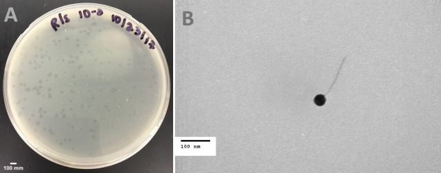 Fig 1
Fig 1Peer Reviews
No public reviews on file for this paper yet. If you reviewed it on a platform where reviews are public (OpenReview, ICLR, NeurIPS, ICML), you can paste yours below so the community can read it here.
Videos
No videos yet. Explain this paper in a talk, walkthrough, or lecture? Add one.
Taxonomy
TopicsBacteriophages and microbial interactions · Genomics and Phylogenetic Studies · Mycobacterium research and diagnosis
ANNOUNCEMENT
The characterization of novel Mycobacterium bacteriophages may advance the development of therapeutics for Mycobacterium leprae and Mycobacterium tuberculosis infections (1). Here, we present the characterization and genome sequence of an actinophage, Rummer, infecting Mycobacterium smegmatis.
Rummer was isolated in September 2017 from soil collected on the campus of Merrimack College in North Andover, MA (global positioning system coordinates 42.6686 N, 71.1216 W) using standard procedures (2). Briefly, the soil was incubated in liquid 7H9 media for 2 hours at 37°C, with shaking, and filtered through a 0.2 µm filter. The filtrate was added to a liquid culture containing late log-phase Mycobacterium smegmatis mc^2^155 and incubated for 24 hours at 37°C, with shaking. The liquid fraction of the overnight culture was filtered (0.2 µm pore size) and the filtrate was spotted on a lawn of M. smegmatis in top agar. Rummer was plaque-purified from the filtrate via three rounds of plating, with plates incubated at 37°C for 24 hours. Rummer plaques were turbid in appearance and approximately 2 mm in diameter (Fig. 1A). Analysis of phage particles by negative-stain transmission electron microscopy revealed that Rummer has a siphovirus morphology with a capsid diameter of 50 nm and a tail length of approximately 160 nm (n = 1, Fig. 1B).
Characterization of Rummer. (A) Plaque morphology. Plaques produced on a lawn of M. smegmatis PYCa medium on a petri dish (90 mm diameter). Plaques are with a clear center surrounded by a turbid edge. (B) Negative-stain (1% uranyl acetate) transmission electron micrograph of Rummer. Phage particles were analyzed using JEOL JEM-1400 Transmission Electron Microscope at an accelerating voltage of 80 kV. Rummer has a siphoviral morphology with a capsid diameter of 50 nm and a tail length of 160 nm (n = 1).
Genomic DNA extraction was performed from filtered high-titer lysates using the Promega Wizard DNA kit. A sequencing library was prepared using the NEBNext Ultra II FS kit and the genome was sequenced using an Illumina MiSeq (v3 reagents), yielding 722,374 150-base single-end reads and 2,012-fold coverage. Raw reads were assembled and checked for completeness using Newbler v2.9 and Consed v29 (3, 4). The resulting genome has a 10-base 3´ single-stranded overhang (CGGGTGGTAA), a G + C content of 64.1%, and a length of 50,908 base pair. An automated annotation was generated using DNA Master (cobamide2.bio.pitt.edu; v5.23.6, build 2705, 24 October 2021), Glimmer (v3.02b) (5), and GeneMark (6), and start sites manually refined utilizing Starterator (http://phages.wustl.edu/starterator/) and Phamerator (7). Functions for putative genes were assigned using the PECAAN (discover.kbrinsgd.org) platform and based on Phagesdb Function Frequency, Phagesdb BLAST, and NCBI BLAST (Blastp from NCBI blast version 2.13.0+) (8) and HHPRED (against the PDB mmCIF70, Pfam-A, and NCBI Conserved Domain databases) (9). Three tRNAs were identified using ARAGORN (v1.2.41) (10) and tRNAscan-SE (v2.0) (11). Default parameters were used for all programs.
Eighty-nine putative genes were identified as protein-coding genes, of which 43 could be assigned putative functions. Based on gene content similarity of >35% to other phages in the Actinobacteriophage Database, PhagesDB (12, 13), Rummer was assigned to cluster A, subcluster A3, which all share the following characteristics: 3´ single-stranded genome ends, structural proteins encoded located on one-half of the genome and transcribed in the forward direction while DNA metabolism functions are encoded on the other half and transcribed in the reverse direction. A noteworthy feature of Rummer is that it is in a subset of phages A3 subcluster phages (13%) that encode three tRNAs. As with a majority of cluster A phages, Rummer encodes putative immunity repressor and integrase functions, suggesting it is a temperate phage.
The reference list from the paper itself. Each links out to its DOI / PubMed record.
- 1Hatfull GF. 2018. Mycobacteriophages. Microbiol Spectr 6. doi:10.1128/microbiolspec.GPP 3-0026-2018 PMC 628202530291704 · doi ↗ · pubmed ↗
- 2Poxleitner M, Pope W, Jacobs-Sera D, Sivanathan V, Hatfull GF. 2018. HHMI SEA-PHAGES phage discovery guide. https://seaphagesphagediscoveryguide.helpdocsonline.com/home.
- 3Gordon D, Green P. 2013. Consed: a graphical editor for next-generation sequencing. Bioinformatics 29:2936–2937. doi:10.1093/bioinformatics/btt 51523995391 PMC 3810858 · doi ↗ · pubmed ↗
- 4Russell DA. 2018. Sequencing, assembling, and finishing complete bacteriophage genomes. Methods Mol Biol 1681:109–125. doi:10.1007/978-1-4939-7343-9_929134591 · doi ↗ · pubmed ↗
- 5Delcher AL, Bratke KA, Powers EC, Salzberg SL. 2007. Identifying bacterial genes and endosymbiont DNA with Glimmer. Bioinformatics 23:673–679. doi:10.1093/bioinformatics/btm 00917237039 PMC 2387122 · doi ↗ · pubmed ↗
- 6Besemer J, Borodovsky M. 2005. Gene Mark: web software for gene finding in prokaryotes, eukaryotes and viruses. Nucleic Acids Res 33:W 451–W 454. doi:10.1093/nar/gki 48715980510 PMC 1160247 · doi ↗ · pubmed ↗
- 7Cresawn SG, Bogel M, Day N, Jacobs-Sera D, Hendrix RW, Hatfull GF. 2011. Phamerator: a bioinformatic tool for comparative bacteriophage genomics. BMC Bioinformatics 12:2105–2112 doi:10.1186/1471-2105-12-395PMC 323361221991981 · doi ↗ · pubmed ↗
- 8Altschul SF, Gish W, Miller W, Myers EW, Lipman DJ. 1990. Basic local alignment search tool. J Mol Biol 215:403–410. doi:10.1016/S 0022-2836(05)80360-22231712 · doi ↗ · pubmed ↗