Down-sampling in diffusion MRI: a bundle-specific DTI and NODDI study
Federico Spagnolo, Susanna Gobbi, Enikő Zsoldos, Manon Edde, Matthias Weigel, Cristina Granziera, Maxime Descoteaux, Muhamed Barakovic, Stefano Magon

TL;DR
This study shows that reducing the number of measurements in diffusion MRI scans can significantly cut acquisition time without greatly affecting key brain imaging metrics.
Contribution
The study introduces a method to down-sample diffusion MRI data while maintaining clinically relevant DTI and NODDI metrics for white matter bundles.
Findings
DTI and NODDI metrics remained comparable at 30% sampling, with FA and MD showing median L1 distances of up to 3.92% and 4.31%, respectively.
At 50% sampling, ICvf showed a median L1 distance of 3.95%, and white matter volume differences were minimal at 2.09%.
Reducing dMRI volumes by 30% using specific gradient directions per shell can maintain accuracy while cutting scan time.
Abstract
Multi-shell diffusion Magnetic Resonance Imaging (dMRI) data has been widely used to characterise white matter microstructure in several neurodegenerative diseases. The lack of standardised dMRI protocols often implies the acquisition of redundant measurements, resulting in prolonged acquisition times. In this study, we investigate the impact of the number of gradient directions on Diffusion Tensor Imaging (DTI) and on Neurite Orientation Dispersion and Density Imaging (NODDI) metrics. Data from 124 healthy controls collected in three different longitudinal studies were included. Using an in-house algorithm, we reduced the number of gradient directions in each data shell. We estimated DTI and NODDI measures on six white matter bundles clinically relevant for neurodegenerative diseases. Fractional Anisotropy (FA) measures on bundles where data were sampled at the 30% rate, showed a…
Genes, proteins, chemicals, diseases, species, mutations and cell lines named across the full text — each resolved to its canonical identifier and authoritative record.
Click any figure to enlarge with its caption.
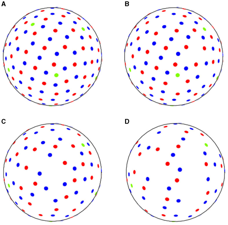 Figure 1
 Figure 2
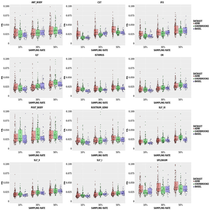 Figure 3
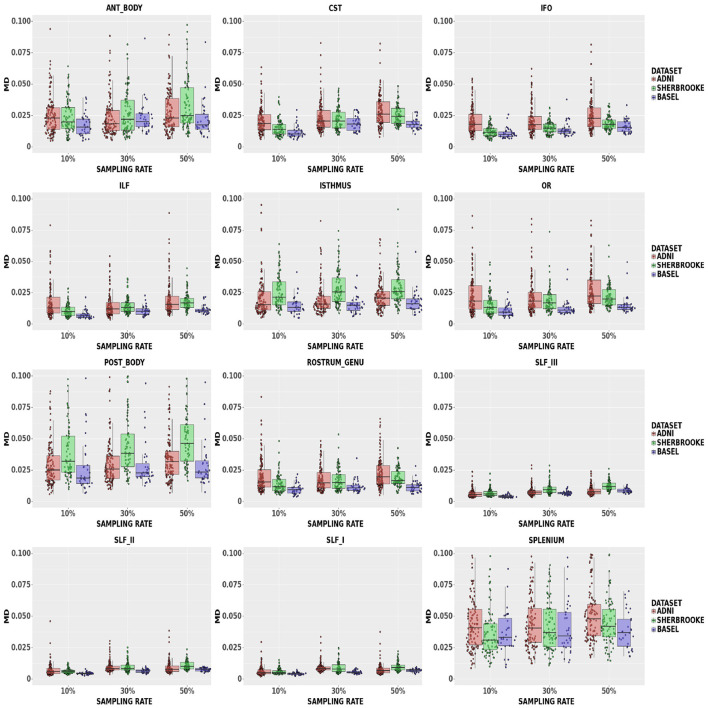 Figure 4
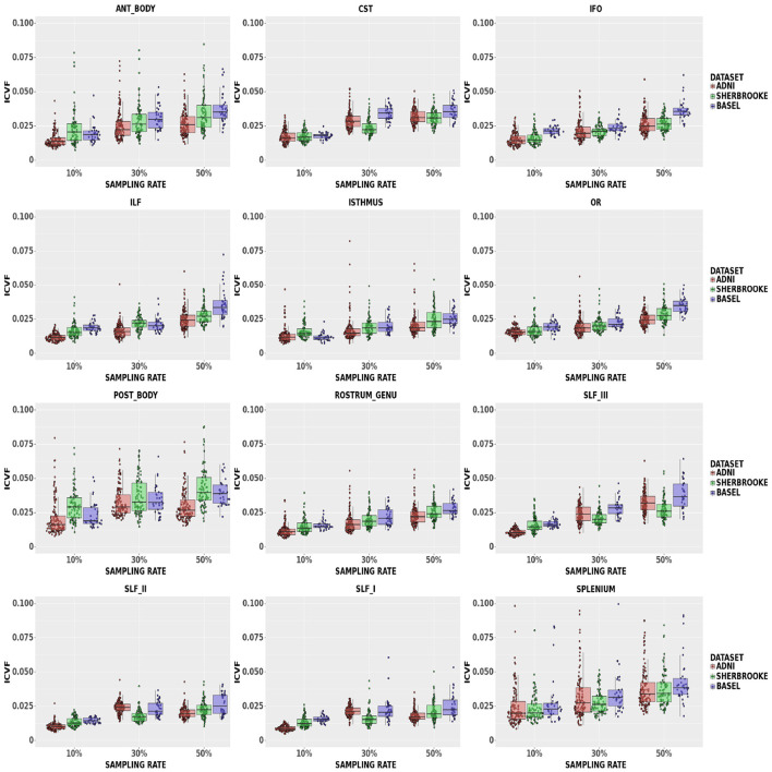 Figure 5
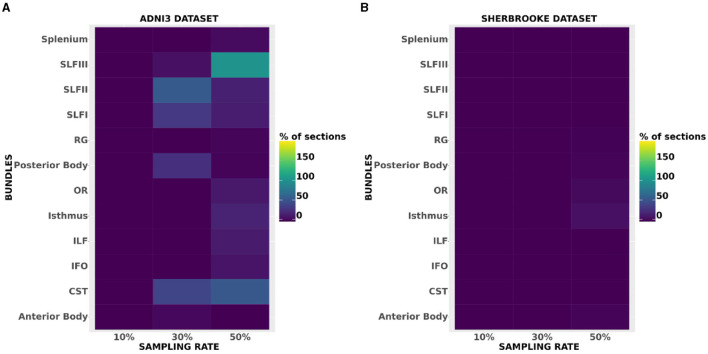 Figure 6
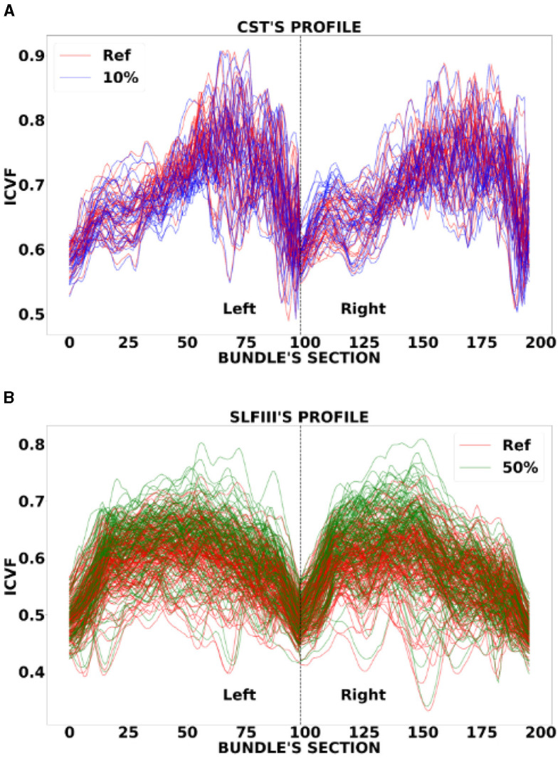 Figure 7
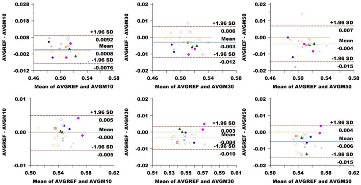 Figure 8
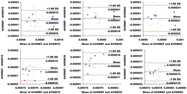 Figure 9
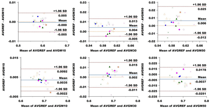 Figure 10
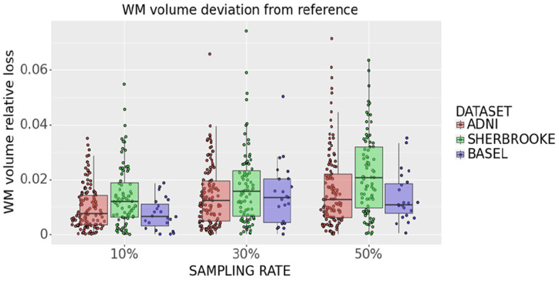 Figure 11
Peer Reviews
No public reviews on file for this paper yet. If you reviewed it on a platform where reviews are public (OpenReview, ICLR, NeurIPS, ICML), you can paste yours below so the community can read it here.
Videos
No videos yet. Explain this paper in a talk, walkthrough, or lecture? Add one.
Taxonomy
TopicsAdvanced Neuroimaging Techniques and Applications · MRI in cancer diagnosis · Advanced MRI Techniques and Applications