A Unique Surgical Case of Mixed Metaplastic Breast Carcinoma With Heterologous Mesenchymal Differentiation and Conventional Adenocarcinomatous Elements
Yoshiiku Okanemasa, Akihiro Shioya, Motona Kumagai, Mao Takata, Yumi Tsubata, Jia Han, Toshie Terauchi, Emi Morioka, Masafumi Inokuchi, Sohsuke Yamada

TL;DR
This paper presents a rare case of mixed metaplastic breast carcinoma with both mesenchymal and adenocarcinomatous elements, emphasizing the importance of accurate diagnosis for better patient outcomes.
Contribution
The paper reports the first case of mixed MBC with detailed FNA cytomorphologic findings and heterologous mesenchymal differentiation.
Findings
Mixed MBC with heterologous mesenchymal differentiation and conventional adenocarcinomatous elements was diagnosed via FNA and confirmed post-surgery.
Cytological features included cohesive clusters of atypical spindled and epithelioid cells with hyperchromatic nuclei and mitotic figures.
Immunohistochemistry confirmed sarcomatous tumor cells positive for vimentin, α-smooth muscle actin, and epithelial membrane antigen.
Abstract
Metaplastic breast carcinoma (MBC) is very rare among all invasive breast carcinomas, accounting for less than 1.0% of them. MBCs are classified into five subtypes, including mixed MBC - where the mix might be multiple metaplastic elements or a mixture of epithelial and mesenchymal elements. Overall survival for mixed MBC tends to correlate with a significantly worse outcome. Therefore, an early accurate diagnosis and surgical treatment for mixed MBCs must allow for an improved quality of life and better prognosis. However, there have not been many recently published papers describing the detailed cytological features of mixed MBCs on fine-needle aspiration (FNA) specimens. A 60-year-old female presented with a history of a hard breast mass on the left lateral side, showing an ill-defined and marginally enhanced tumor nodule on magnetic resonance imaging. The cytologic specimens of FNA…
Genes, proteins, chemicals, diseases, species, mutations and cell lines named across the full text — each resolved to its canonical identifier and authoritative record.
Click any figure to enlarge with its caption.
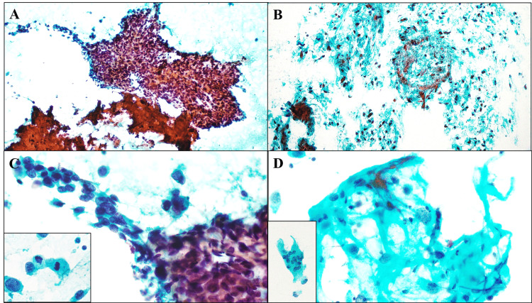 Figure 1
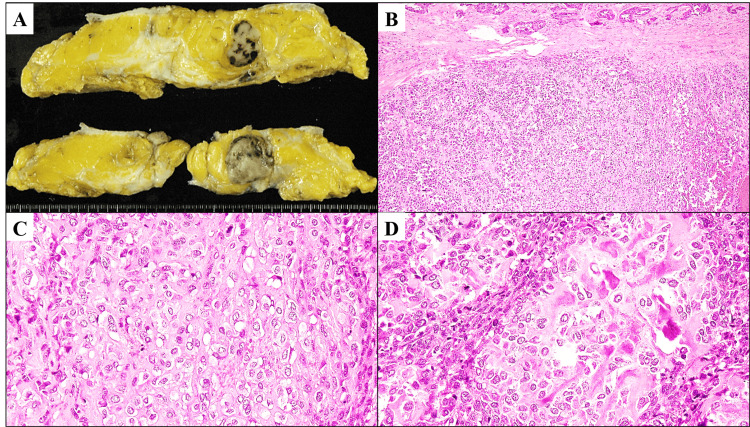 Figure 2
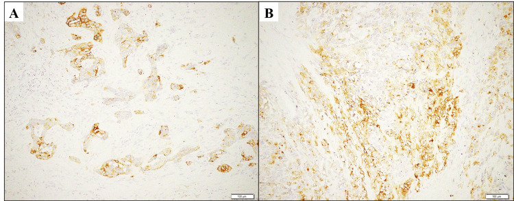 Figure 3
Peer Reviews
No public reviews on file for this paper yet. If you reviewed it on a platform where reviews are public (OpenReview, ICLR, NeurIPS, ICML), you can paste yours below so the community can read it here.
Videos
No videos yet. Explain this paper in a talk, walkthrough, or lecture? Add one.
Taxonomy
TopicsBreast Lesions and Carcinomas · Cancer and Skin Lesions · Metastasis and carcinoma case studies