Usefulness of a novel drill dilator in removing a migrated biliary plastic internal stent
Takafumi Yanaidani, Tomoaki Matsumori, Masataka Yokode, Yuya Muramoto, Masahiro Shiokawa, Norimitsu Uza, Hiroshi Seno

Abstract
Genes, proteins, chemicals, diseases, species, mutations and cell lines named across the full text — each resolved to its canonical identifier and authoritative record.
Click any figure to enlarge with its caption.
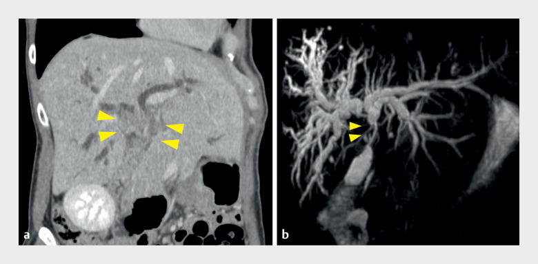 Fig. 1
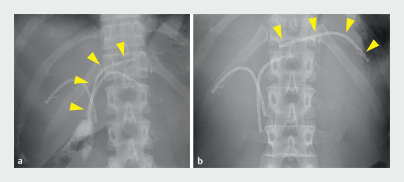 Fig. 2
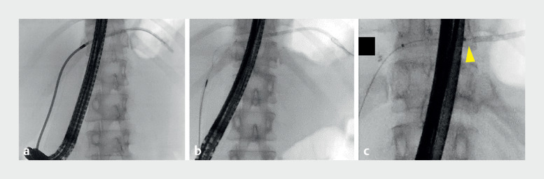 Fig. 3
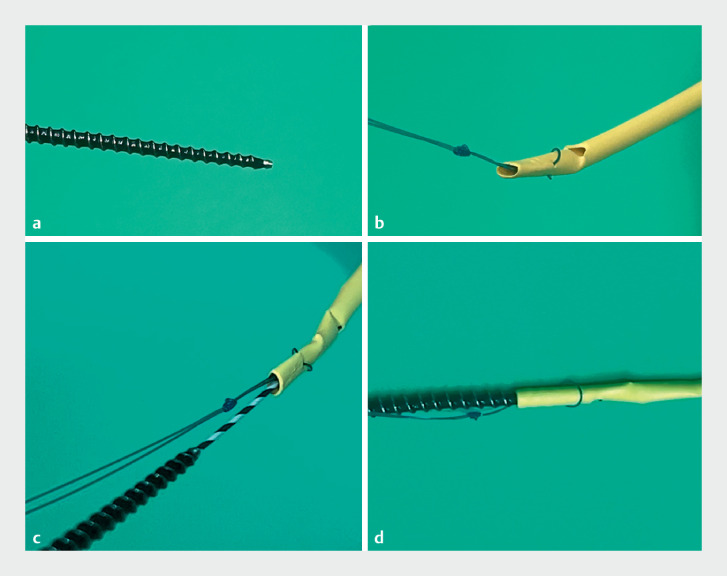 Fig. 4
Peer Reviews
No public reviews on file for this paper yet. If you reviewed it on a platform where reviews are public (OpenReview, ICLR, NeurIPS, ICML), you can paste yours below so the community can read it here.
Videos
No videos yet. Explain this paper in a talk, walkthrough, or lecture? Add one.
Taxonomy
TopicsGallbladder and Bile Duct Disorders · Esophageal and GI Pathology · Pediatric Hepatobiliary Diseases and Treatments
A 45-year-old woman referred to hospital for preoperative evaluation of perihilar cholangiocarcinoma ( Fig. 1 ) had multiple biliary plastic internal stents placed previously ( Fig. 2 a ). Endoscopic retrograde cholangiography (ERC) was performed to evaluate tumor extent and replace the plastic internal stents. ERC revealed that a nylon-threaded plastic stent (Through & Pass, 7 Fr, 9 cm; Gadelius Medical, Tokyo, Japan) placed in the left hepatic duct (B2) had migrated into the peripheral bile duct ( Fig. 2 b ). The nylon threads had entered the common bile duct and the migrated plastic stent could not be removed by pulling them.
Imaging from the previous hospital. a Computed tomography showed cholangiocarcinoma located in the perihilar bile duct (yellow arrows), with dilated intrahepatic bile ducts. b Magnetic resonance cholangiopancreatography revealed a hilar biliary stricture (yellow arrows) and dilated intrahepatic ducts.
Fluoroscopic images. a Multiple biliary plastic stents were placed at another hospital. b One plastic stent had migrated into the peripheral bile duct; yellow arrowheads indicate the stent at B2.
Attempts to remove the stent using a stent retriever (Soehendra Stent Retriever; Cook Medical Japan, Tokyo, Japan) and snare catheter (SD-5U-1; Olympus, Tokyo, Japan), failed, breaking and deforming the proximal stent tip in the process ( Fig. 3 ). A guidewire was inserted into the migrated internal stent in an attempt to insert a balloon catheter (REN; Kaneka Co., Inc., Osaka, Japan) into the stent along the guidewire; however, it could not be inserted because of the broken stent tip. Finally, we used a novel 7-Fr drill dilator with a tapered screw-shaped tip (Tornus ES; Olympus) ( Fig. 4 a , Video 1 ). Using pushing and clockwise rotation, the dilator was passed through the biliary stricture and easily inserted into the broken stent tip along the guidewire, facilitating successful removal of the stent ( Fig. 4 b–d ). New plastic internal stents were placed after tumor evaluation.
Fluoroscopic images of conventional stent removal methods for a migrated biliary plastic stent. a A guidewire was inserted into the stent and a stent retriever was inserted along the guidewire. b A snare catheter was used to grasp the migrated stent. c Insertion of a balloon catheter (yellow arrowhead: balloon catheter tip) into the stent was challenging due to the broken stent tip.
Removal of the biliary plastic stent using a novel drill dilator. a Tip shape of the novel drill dilator. b Broken and deformed proximal tip of the biliary plastic stent. c The guidewire was passed through the stent, and the novel device proceeded along the guidewire. d The novel device was inserted into the broken proximal tip of the plastic stent, facilitating successful removal of the stent.
Insertion of the novel drill dilator into the migrated biliary plastic stent and successful stent removal.Video 1
Although there are several methods for removal of biliary stents 1 2 , some cases remain challenging when the stent migrates into the peripheral bile duct. Use of this novel dilator for endoscopic procedures, such as the removal of biliary stents placed across the duodenal papilla, has been reported 3 4 5 . This novel drill dilator has a tapered, sharp, corkscrew tip allowing passage through the biliary stricture and firm grasp of the plastic stent, making it useful for removing migrated biliary internal stents in technically challenging cases.
Endoscopy_UCTN_Code_TTT_1AR_2AZ
The reference list from the paper itself. Each links out to its DOI / PubMed record.
- 1Yalaka RR Kedarisetty CK Narayankar SMA novel technique of migrated biliary stent retrieval Endoscopy 202254 E 522E 52310.1055/a-1662-392034695850 · doi ↗ · pubmed ↗
- 2Calcara C Broglia L Comi G Plastic biliary stent migration during multiple stents placement and successful endoscopic removal using intra-stent balloon inflation technique: a case report and literature review Am J Case Rep 201617656926847214 10.12659/AJCR.896076 PMC 4745661 · doi ↗ · pubmed ↗
- 3Shiomi H Nakano R Iijima H Recanalization using a novel drill-shaped dilator for a severe pancreatic duct stricture and impacted pancreatic duct stone Endoscopy 202355 E 351E 35310.1055/a-1986-729536646122 PMC 9842438 · doi ↗ · pubmed ↗
- 4Yamada M Hara K Haba S Endoscopic ultrasound-guided hepaticogastrostomy using a novel drill dilator Endoscopy 202254 E 856E 85710.1055/a-1838-368235636451 PMC 9735340 · doi ↗ · pubmed ↗
- 5Ishida Y Koga T Hirai F Successful removal of a migrated biliary plastic stent using a novel spiral dilator Endoscopy 202355 E 804E 80510.1055/a-2098-098237321269 PMC 10270752 · doi ↗ · pubmed ↗